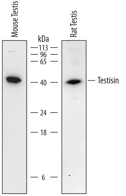
Detection of Mouse and Rat Testisin/Prss21 by Western Blot. Western blot shows lysates of mouse testis tissue and rat testis tissue. PVDF membrane was probed with 0.5 µg/mL of Goat Anti-Mouse Testisin/Prss21 Antigen Affinity-purified Polyclonal Antibody (Catalog # AF6820) followed by HRP-conjugated Anti-Goat IgG Secondary Antibody (Catalog # HAF017). A specific band was detected for Testisin/Prss21 at approximately 40 kDa (as indicated). This experiment was conducted under reducing conditions and using Immunoblot Buffer Group 1.
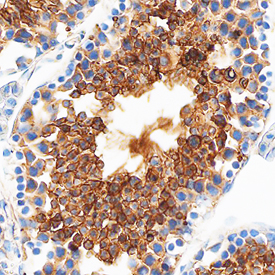
Testisin/Prss21 in Mouse Testis. Testisin/Prss21 was detected in perfusion fixed frozen sections of mouse testis using Goat Anti-Mouse Testisin/Prss21 Antigen Affinity-purified Polyclonal Antibody (Catalog # AF6820) at 1.7 µg/mL overnight at 4 °C. Tissue was stained using the Anti-Goat HRP-DAB Cell & Tissue Staining Kit (brown; Catalog # CTS008) and counterstained with hematoxylin (blue). Specific staining was localized to the plasma membranes of spermatocytes. View our protocol for Chromogenic IHC Staining of Frozen Tissue Sections. This application has not been tested in rat samples.