Anti-CAPZA1 antibody [EPR11210]
| Name | Anti-CAPZA1 antibody [EPR11210] |
|---|---|
| Supplier | Abcam |
| Catalog | ab166892 |
| Prices | $357.00 |
| Sizes | 100 µl |
| Host | Rabbit |
| Clonality | Monoclonal |
| Isotype | IgG |
| Clone | EPR11210 |
| Applications | WB IHC-P IP FC |
| Species Reactivities | Mouse, Rat, Human |
| Antigen | Recombinant fragment corresponding to residues in Human CAPZA1 (UniProt ID: P52907) |
| Description | Rabbit Monoclonal |
| Gene | CAPZA1 |
| Conjugate | Unconjugated |
| Supplier Page | Shop |
Product images
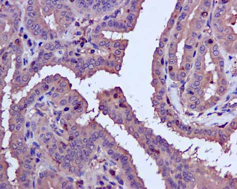 ab166892 showing +ve staining in Human thyroid gland carcinoma.
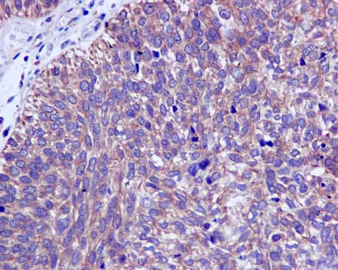 ab166892 showing +ve staining in Human urinary bladder carcinoma.
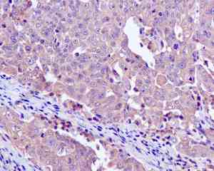
 ab166892 showing +ve staining in Human colonic adenocarcinoma.
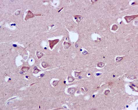 ab166892 showing +ve staining in Human normal brain.
![All lanes : Anti-CAPZA1 antibody [EPR11210] (ab166892) at 1/1000 dilutionLane 1 : HeLa cell lysateLane 2 : Jurkat cell lysateLane 3 : 293T cell lysateLane 4 : HepG2 cell lysateLysates/proteins at 10 µg per lane.SecondaryHRP labelled goat anti-rabbit at 1/2000 dilution](http://www.bioprodhub.com/system/product_images/ab_products/2/sub_1/20355_CAPZA1-Primary-antibodies-ab166892-1.jpg) All lanes : Anti-CAPZA1 antibody [EPR11210] (ab166892) at 1/1000 dilutionLane 1 : HeLa cell lysateLane 2 : Jurkat cell lysateLane 3 : 293T cell lysateLane 4 : HepG2 cell lysateLysates/proteins at 10 µg per lane.SecondaryHRP labelled goat anti-rabbit at 1/2000 dilution
All lanes : Anti-CAPZA1 antibody [EPR11210] (ab166892) at 1/1000 dilutionLane 1 : HeLa cell lysateLane 2 : Jurkat cell lysateLane 3 : 293T cell lysateLane 4 : HepG2 cell lysateLysates/proteins at 10 µg per lane.SecondaryHRP labelled goat anti-rabbit at 1/2000 dilution
 ab166892, at 1/1000 dilution, staining CAPZA1 in HepG2 cell lysate immunoprecipitated using ab166892 at 1/10.
 Immunohistochemical analysis of paraffin embedded Human cervical carcinoma tissue labeling CAPZA1 with ab166892 at 1/100 dilution.
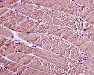 Immunohistochemical analysis of paraffin embedded Human muscle tissue labeling CAPZA1 with ab166892 at 1/100 dilution.
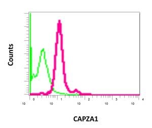 Flow cytometric analysis of permeabilized Jurkat cells using ab166892 at a dilution of 1/10 (red) compared to a rabbit IgG (negative) (green).