Anti-CCNI2 antibody
| Name | Anti-CCNI2 antibody |
|---|---|
| Supplier | Abcam |
| Catalog | ab170531 |
| Prices | $370.00 |
| Sizes | 400 µl |
| Host | Rabbit |
| Clonality | Polyclonal |
| Isotype | IgG |
| Applications | ICC/IF ICC/IF WB IHC-P |
| Species Reactivities | Human |
| Antigen | Synthetic peptide within Human CCNI2 aa 92-121 (internal sequence) conjugated to Keyhole Limpet Haemocyanin (KLH) |
| Description | Rabbit Polyclonal |
| Gene | CCNI2 |
| Conjugate | Unconjugated |
| Supplier Page | Shop |
Product images
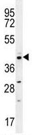 Anti-CCNI2 antibody (ab170531) + HepG2 cell lysate at 35 µgdeveloped using the ECL technique
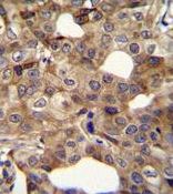 Immunohistochemistry (Formalin/PFA-fixed paraffin-embedded sections) analysis of human breast carcinoma tissue labelling CCNI2 with ab170531. A peroxidase-conjugated anti-rabbit IgG was used as the secondary antibody, followed by DAB staining.
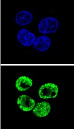 Immunocytochemistry/Immunofluorescence analysis of HepG2 cells labeling CCNI2 (green) with ab170531. An Alexa Fluor® 488-conjugated goat anti-rabbit lgG was used as the secondary antibody. DAPI was used to stain the cell nuclei (blue).