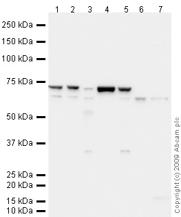
All lanes : Anti-CD80 antibody (ab64116) at 1 µg/mlLane 1 : Spleen (Mouse) Tissue Lysate Lane 2 : Thymus (Mouse) Tissue Lysate Lane 3 : Brain (Mouse) Tissue Lysate Lane 4 : Skeletal Muscle (Mouse) Tissue Lysate Lane 5 : Spinal Cord (Mouse) Tissue Lysate Lane 6 : EL4 (Mouse lymphoma cell line) Whole Cell Lysate (ab7183)Lane 7 : RAW 264.7 (Mouse leukaemic monocyte macrophage cell line) Whole Cell Lysate Lysates/proteins at 10 µg per lane.SecondaryGoat polyclonal to Rabbit IgG - H&L - Pre-Adsorbed (HRP) (ab65484) at 1/3000 dilutiondeveloped using the ECL techniquePerformed under reducing conditions.
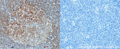
ab64116 (1:40) staining CD80 in paraffin-embedded mouse lymph node tissue (left panel) using an automated system (Ventana Discovery). Right-hand panel shows negative control (no primary antibody). Using this protocol there is strong membrane staining, of activated B cells in the germinal centres. What appear to be scattered activated B cells or possibly plasma cells in the interfollicular areas of the lymph node and blood vessels.Sections were rehydrated and antigen retrieved in CC1 Cell Conditioning Buffer using Ventana Extended Retrieval programme. Slides were blocked in 3% H2O2 / 4 min / 37°C and incubated with ab64116 (1:40 dilution / 1 hour / 37°C). Sections then blocked (4mins / 37°C) and incubated with Dako swine anti-rabbit antibody (1:50, 28 min / 37°C). Staining was amplified and detected by incubation with Ventana Streptavidin ABC system (16 min / 37°C) and Ventana DAB map reagent (8 min / 37°C). Slides were counterstained with Haematoxylin and coverslipped in DPX.For man