![Anti-CDT1 antibody [EPR17891] (ab202067) at 1/10000 dilution + HeLa (Human epithelial cells from cervix adenocarcinoma) cell lysate at 20 µgSecondaryGoat Anti-Rabbit IgG, (H+L), Peroxidase conjugated at 1/1000 dilution](http://www.bioprodhub.com/system/product_images/ab_products/2/sub_1/27158_ab202067-244479-ab202067WBa.jpg)
Anti-CDT1 antibody [EPR17891] (ab202067) at 1/10000 dilution + HeLa (Human epithelial cells from cervix adenocarcinoma) cell lysate at 20 µgSecondaryGoat Anti-Rabbit IgG, (H+L), Peroxidase conjugated at 1/1000 dilution
![Anti-CDT1 antibody [EPR17891] (ab202067) at 1/2000 dilution + HEK-293 (Human epithelial cells from embryonic kidney) cell lysate at 20 µgSecondaryGoat Anti-Rabbit IgG, (H+L), Peroxidase conjugated at 1/1000 dilution](http://www.bioprodhub.com/system/product_images/ab_products/2/sub_1/27159_ab202067-244478-ab202067WBb.jpg)
Anti-CDT1 antibody [EPR17891] (ab202067) at 1/2000 dilution + HEK-293 (Human epithelial cells from embryonic kidney) cell lysate at 20 µgSecondaryGoat Anti-Rabbit IgG, (H+L), Peroxidase conjugated at 1/1000 dilution
![Anti-CDT1 antibody [EPR17891] (ab202067) at 1/1000 dilution + Human fetal lung lysate at 10 µgSecondaryAnti-Rabbit IgG (HRP), specific to the non-reduced form of IgG at 1/1000 dilution](http://www.bioprodhub.com/system/product_images/ab_products/2/sub_1/27160_ab202067-244477-ab202067WBc.jpg)
Anti-CDT1 antibody [EPR17891] (ab202067) at 1/1000 dilution + Human fetal lung lysate at 10 µgSecondaryAnti-Rabbit IgG (HRP), specific to the non-reduced form of IgG at 1/1000 dilution
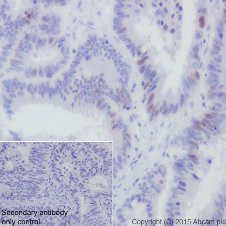
Immunohistochemical analysis of paraffin-embedded Human colonic carcinoma tissue labeling CDT1 with ab202067 at 1/100 dilution, followed by Goat Anti-Rabbit IgG H&L (HRP) (ab97051) secondary antibody at 1/500 dilution.Nuclear staining on Human colonic carcinoma tissue is observed.Counter stained with Hematoxylin.Secondary antibody only control: Used PBS instead of primary antibody, secondary antibody is Goat Anti-Rabbit IgG H&L (HRP) (ab97051) at 1/500 dilution.
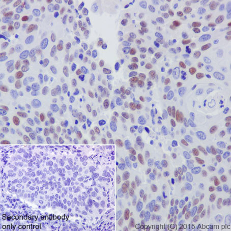
Immunohistochemical analysis of paraffin-embedded Human cervix carcinoma tissue labeling CDT1 with ab202067 at 1/100 dilution, followed by Goat Anti-Rabbit IgG H&L (HRP) (ab97051) secondary antibody at 1/500 dilution. Nuclear staining on Human cervix carcinoma tissue is observed.Counter stained with Hematoxylin.Secondary antibody only control: Used PBS instead of primary antibody, secondary antibody is Goat Anti-Rabbit IgG H&L (HRP) (ab97051) at 1/500 dilution.
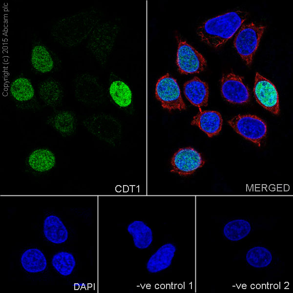
Immunofluorescent analysis of 4% paraformaldehyde-fixed, 0.1% Triton X-100 permeabilized HeLa (Human epithelial cells from cervix adenocarcinoma) cells labeling CDT1 with ab202067 at 1/100 dilution, followed by Goat anti-rabbit IgG (Alexa Fluor® 488) (ab150077) secondary antibody at 1/500 dilution (green).Confocal image showing nuclear staining on HeLa cell line.The nuclear counter stain is DAPI (blue).Tubulin is detected with ab7291 (anti-Tubulin mouse mAb) at 1/1000 dilution and ab150120 (AlexaFluor®594 Goat anti-Mouse secondary) at 1/500 dilution (red).The negative controls are as follows:-ve control 1: ab202067 at 1/100 dilution followed by ab150120 (AlexaFluor®594 Goat anti-Mouse secondary) at 1/500 dilution.-ve control 2: ab7291 (anti-Tubulin mouse mAb) at 1/1000 dilution followed by ab150077 (Alexa Fluor®488 Goat Anti-Rabbit IgG H&L) at 1/500 dilution.
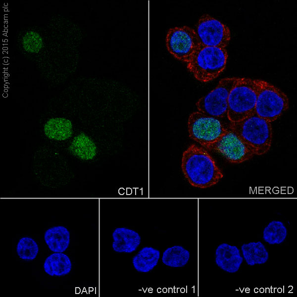
Immunofluorescent analysis of 4% paraformaldehyde-fixed, 0.1% Triton X-100 permeabilized HT-29 (Human colorectal adenocarcinoma cells) cells labeling CDT1 with ab202067 at 1/100 dilution, followed by Goat anti-rabbit IgG (Alexa Fluor® 488) (ab150077) secondary antibody at 1/500 dilution (green).Confocal image showing nuclear staining on T-29 cell line.The nuclear counter stain is DAPI (blue).Tubulin is detected with ab7291 (anti-Tubulin mouse mAb) at 1/1000 dilution and ab150120 (AlexaFluor®594 Goat anti-Mouse secondary) at 1/500 dilution (red).The negative controls are as follows:-ve control 1: ab202067 at 1/100 dilution followed by ab150120 (AlexaFluor®594 Goat anti-Mouse secondary) at 1/500 dilution.-ve control 2: ab7291 (anti-Tubulin mouse mAb) at 1/1000 dilution followed by ab150077 (Alexa Fluor®488 Goat Anti-Rabbit IgG H&L) at 1/500 dilution.
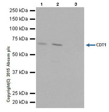
CDT1 was immunoprecipitated from 1mg of HEK-293 (Human epithelial cells from embryonic kidney) whole cell lysate with ab202067 at 1/30 dilution.Western blot was performed from the immunoprecipitate using ab202067 at 1/1000 dilution.Anti-Rabbit IgG (HRP), specific to the non-reduced form of IgG was used as secondary antibody at 1/1500 dilution.Lane 1: HEK-293 whole cell lysate 10 µg (Input).Lane 2: ab202067 IP in HEK-293 whole cell lysate.Lane 3: Rabbit monoclonal IgG (ab172730) instead of ab202067 in HEK-293 whole cell lysate.Blocking and dilution buffer and concentration: 5% NFDM/TBST.Exposure time: 5 seconds.