
Immunocytochemistry with anti-cleaved PARP antibody conjugated to Alexa Fluor® 488.HeLa cells were vehicle-treated (panels A-C) or treated with 1 µM staurosporine for 4 hours (panels D-F), then fixed. Cells were treated with antigen retrieval buffer (100 mM Tris, 5% urea, pH 9.5) for 10 minutes at 95°C, then permeabilized and blocked. Cells were incubated with 1 µg/mL of the cleaved PARP antibody conjugated to Alexa Fluor® 488, then co-stained with the DNA stain DAPI. Images of DAPI signals (A and D), anti-cleaved PARP signal (B and E), and overlays of DAPI (artificially colored red for better contrast) and anti-cleaved PARP (colored green) images (C and F) are shown.
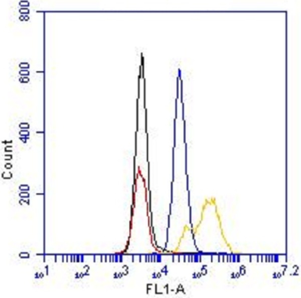
Flow cytometry with anti-cleaved PARP antibody conjugated to Alexa Fluor® 488.Flow cytometric analysis was performed on HeLa vehicle-treated cells and on HeLa cells treated with 1 µM staurosporine for 4 hours. Cells were fixed with paraformaldehyde and permeablized with methanol. HeLa vehicle-treated cells were stained with 1 µg/mL of the cleaved PARP antibody conjugated to Alexa488 (blue) or a negative, nonreactive Alexa Fluor® 488-conjugated control antibody (black). HeLa staurosporine-treated cells were stained with 1 µg/mL of the cleaved PARP antibody conjugated to Alexa Fluor® 488 (yellow) or a negative, nonreactive Alexa Fluor® 488-conjugated control antibody (red). 1% BSA in PBS was used as the blocking reagent for all blocking and antibody incubation steps.