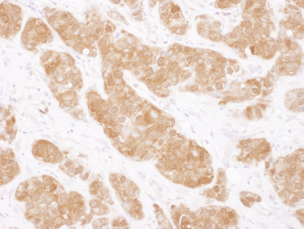
Immunohistochemistry (Formalin/PFA-fixed paraffin-embedded sections) analysis of human breast carcinoma tissue labelling COMT with ab129504 at 1/1000 (1µg/ml). Detection: DAB.

All lanes : Anti-COMT antibody (ab129504) at 0.1 µg/mlLane 1 : Whole cell lysate from HeLa cells at 50 µgLane 2 : Whole cell lysate from HeLa cells at 15 µgLane 3 : Whole cell lysate from 293T cells at 50 µgLane 4 : Whole cell lysate from Jurkat cells at 50 µgdeveloped using the ECL technique
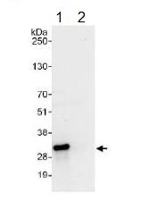
ab129504 at 1 µg/ml staining COMT by WB, following immunoprecipitation of whole cell lysate from HeLa cells. Lane 1; IP using ab129504 at 6 µg/mg lysate. Lane 2; IP using control IgG. 1 mg of lysate was used for IP and 20% of IP was loaded. Detection utilised Chemiluminescence with a 10 second exposure.