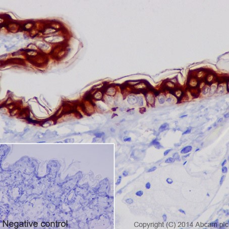
Immunohistochemical analysis of paraffin-embedded mouse skin tissue labeling Cytokeratin 1 with ab185629 at 1/200 dilution, followed by Goat Anti-Rabbit IgG H&L (HRP) (ab97051) secondary antibody at 1/500 dilution. Cytoplasm staining on keratinized epithelium of the mouse skin tissue is observed. Counter stained with Hematoxylin.Negative control: Used PBS instead of primary antibody, secondary antibody is Goat Anti-Rabbit IgG H&L (HRP) (ab97051) at 1/500 dilution.
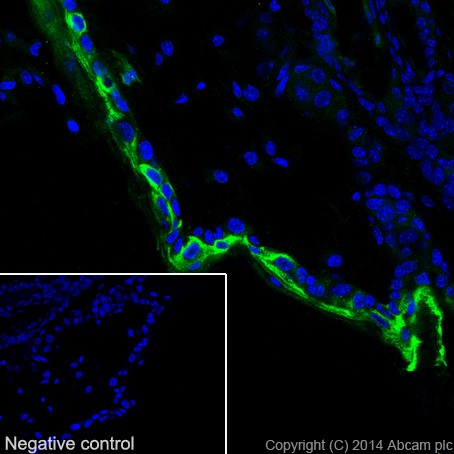
Immunohistochemical analysis of 4% paraformaldehyde-fixed frozen mouse skin tissue labeling Cytokeratin 1 with ab185629 at 1/100 dilution, followed by AlexaFluor®488 Goat anti-Rabbit (ab150077) secondary antibody at 1/500 dilution. Confocal image showing cytoplasm staining on keratinized epithelium of the mouse skin.Negative control: Used PBS instead of primary antibody, secondary antibody is AlexaFluor®488 Goat anti-Rabbit (ab150077) at 1/500 dilution.
![All lanes : Anti-Cytokeratin 1 antibody [EPR17870] (ab185629) at 1/5000 dilutionLane 1 : Mouse skin lysatesLane 2 : Mouse brain lysatesLane 3 : Mouse heart lysatesLane 4 : Mouse kidney lysatesLane 5 : Mouse spleen lysatesLane 6 : RAW 264.7 (Mouse macrophage cells transformed with Abelson murine leukemia virus) whole cell lysateLane 7 : NIH/3T3 (Mouse embyro fibroblast cells) whole cell lysateLysates/proteins at 10 µg per lane.SecondaryGoat Anti-Rabbit IgG, (H+L),Peroxidase conjugated at 1/1000 dilution](http://www.bioprodhub.com/system/product_images/ab_products/2/sub_2/5097_ab185629-237954-185629WBb.jpg)
All lanes : Anti-Cytokeratin 1 antibody [EPR17870] (ab185629) at 1/5000 dilutionLane 1 : Mouse skin lysatesLane 2 : Mouse brain lysatesLane 3 : Mouse heart lysatesLane 4 : Mouse kidney lysatesLane 5 : Mouse spleen lysatesLane 6 : RAW 264.7 (Mouse macrophage cells transformed with Abelson murine leukemia virus) whole cell lysateLane 7 : NIH/3T3 (Mouse embyro fibroblast cells) whole cell lysateLysates/proteins at 10 µg per lane.SecondaryGoat Anti-Rabbit IgG, (H+L),Peroxidase conjugated at 1/1000 dilution

Immunohistochemical analysis of paraffin-embedded mouse kidney tissue labeling Cytokeratin 1 with ab185629 at 1/200 dilution, followed by Goat Anti-Rabbit IgG H&L (HRP) (ab97051) secondary antibody at 1/500 dilution. Negative on mouse kidney tissue. Counter stained with Hematoxylin.
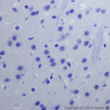
Immunohistochemical analysis of paraffin-embedded mouse brain tissue labeling Cytokeratin 1 with ab185629 at 1/200 dilution, followed by Goat Anti-Rabbit IgG H&L (HRP) (ab97051) secondary antibody at 1/500 dilution. Negative on mouse brain tissue. Counter stained with Hematoxylin.