Rabbit anti-Phospho 53BP1 (S25) Antibody, Affinity Purified
| Name | Rabbit anti-Phospho 53BP1 (S25) Antibody, Affinity Purified |
|---|---|
| Supplier | Sigma-Aldrich |
| Catalog | PLA0126 |
| Prices | $345.00 |
| Sizes | 100 µl |
| Applications | ICC/IF |
| Species Reactivities | Dog, Primate, Ape, Rabbit, Mouse, Chimpanzee, Pig, Bovine, Guinea Pig, Human, Horse, Monkey |
| Gene | TP53BP1 |
| Supplier Page | Shop |
Product images
 Immunohistochemistry
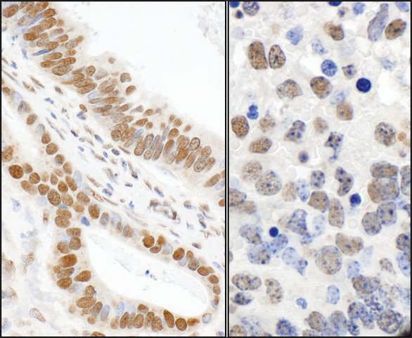
Rabbit Anti-Phospho 53BP1 (S25) Antibody, Affinity Purified: Cat. No. PLA0126: Detection of Human and Mouse Phospho 53BP1 (S25) by Immunohistochemistry. Sample: FFPE sections of Human stomach carcinoma (left) and Mouse teratoma (right). Antibody: Affinity purified Rabbit Anti-Phospho 53bp1 (S25) (Cat. No. PLA0126 ) used at a dilution of 1:1,000 (1 μg/mL). Detection: DAB
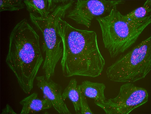 Immunofluorescence: This antibody is qualified for the Proximity Ligation Assay (PLA). The above image shows representative results for PLA using three color fluorescence, including DAPI stained nuclei (blue), phalloidin stained cytoplasmic F-actin (green), and PLA positive signal (red).
 Immunoblotting
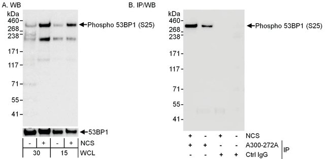
Rabbit Anti-Phospho 53BP1 (S25) Antibody, Affinity Purified: Cat. No. PLA0126: Detection of Phospho 53BP1 (S25) by Western Blot. Samples: Whole cell lysate (30 μg or 15 μg for WB; 1 mg for IP, 20% of IP loaded) from 293T cells that were mock treated or treated with NCS (200 ng/mL; 1 hour). Antibodies: Affinity purified Rabbit Anti-Phospho 53BP1 Antibody PLA0126 at 0.04 μg/mL (A) and 0.1 μg/mL (B) for WB. 53BP1 was also immunoprecipitated using an affinity purified Rabbit Anti-53BP1 Antibody that recognizes a different epitope. Detection: Chemiluminescence with exposure times of 3 seconds (A and B).
 Flow Cytometry
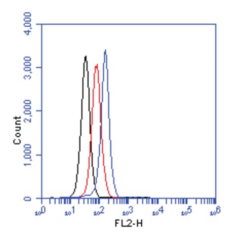
Rabbit Anti-Phospho 53BP1 (S25) Antibody, Affinity Purified: Cat. No. PLA0126: Flow cytometric analysis of phospho 53bp1 (S25). Jurkat cells were treated with neocarzinostatin (NCS) for 1.5 hours. 1 × 10⁶ cells were fixed, permeabilized, and stained with 1.5 μg/mL Anti-phospho 53bp1 (S25) PLA0126 in a 150 mcl reaction. Isotype control (black), untreated (red), NCS treated (blue).