
HeLa cell lysate, Jurkat cell lysate, SH SY5Y cell lysate, MCF7 cell lysate (not shown) and MDA-MB-231 cell lysate were tested using this kit. HeLa cell lysate shown as standard control sample, varying concentration other cell Lysates were analyzed within working range of the assay.
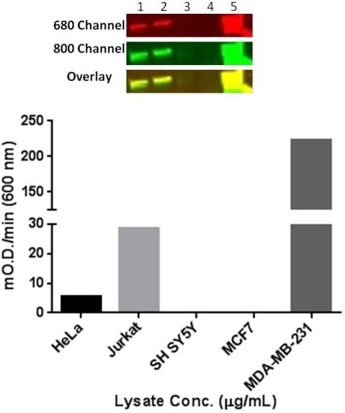
Western blot of cell lysates (top panel): 1) HeLa, 2) Jurkat, 3) Sy5y, 4) MCF7, 5) MDA-MB-231 (10 μg/lane) using primary antibodies: rabbit anti-vimentin (1/1000 dilution) and mouse anti-vimentin (ab8978, 1/1000 dilution); Secondary antibodies used were goat anti‑rabbit 680-RD (Red, 1/10,000) and goat anti‑mouse 800 (Green, 1/10,000). Blot was scanned using a LI‑COR® Odyssey® imager and overlay shows target specificity for total vimentin (yellow). Bar graph (bottom panel) represents relative mOD/min (600 nm) observed for 10 μg/mL cell lysate, except for MDA-MB-231 cell lysate which reports mOD/min (600 nm) for 1 μg/mL.
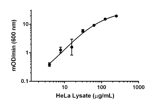 Typical standard curve generated when using the Vimentin Human Profiling ELISA kit.