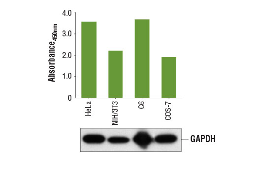
Figure 1. GAPDH protein is detected at varying levels from multiple cell lines using the PathScan® Total GAPDH Sandwich ELISA Kit #7157. The absorbance readings at 450 nm are shown in the top figure, while the corresponding western blots using GAPDH (D16H11) XP® Rabbit mAb #5174 are shown in the bottom figure.
Go to product page
Image may be subject to copyright.