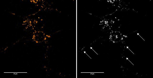
All images provided by Thorsten LauFigure 1: Two neuronal cells stained with 50 μM FFN102 on differentiation day 10. Shown is a sum projection of a confocal z-stack. Accumulation of FFN102 can be observed along the neurites (arrows) and the cell soma (S).
Go to product page
Image may be subject to copyright.