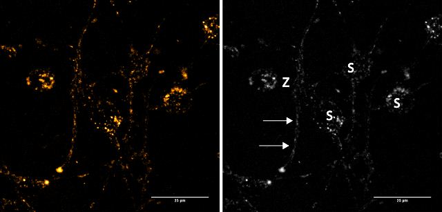
Figure 2a: Images in the first row show a group of neuronal cells stained with 50 μM FFN102 (sum projection of a confocal stack). FFN102 localizes to structures on the cell soma (S) as well as neurites (arrows). Z indicates the area zoomed in for an additional z-stack.
Go to product page
Image may be subject to copyright.