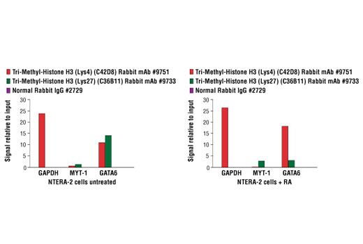
NTERA-2 cells were either untreated (left panel) or treated for 15 days with retinoic acid (RA) to induce differentiation along the neuronal lineage (right panel). Chromatin immunoprecipitations were then performed with cross-linked chromatin from 4 x 10⁶ cells and Tri-Methyl-Histone H3 (Lys4) (C42D8) Rabbit mAb, Tri-Methyl-Histone H3 (Lys27) (C36B11) Rabbit mAb, or 2 μl of Normal Rabbit IgG, using SimpleChIP® Enzymatic Chromatin IP Kit (Magnetic Beads) #9003. The enriched DNA was quantified by real-time PCR using SimpleChIP® Human GAPDH Exon 1 Primers #5516, SimpleChIP® Human MYT-1 Exon 1 Primers #4493, and SimpleChIP® Human GATA6 Promoter Primers #5550. The amount of immunoprecipitated DNA in each sample is normalized for enrichment of total histone H3 and represented as signal relative to the total amount of input chromatin, which is equivalent to one. Note the loss of tri-methyl histone H3 Lys27 on the GATA6 promoter as it is activated during NTERA-2 cell differentiatiation.
Go to product page
Image may be subject to copyright.