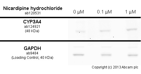
HepG2 cells were incubated at 37°C for 48h with vehicle control (0 µM) and different concentrations of nicardipine hydrochloride (ab120531) in DMSO. Increased expression of cytochrome P450 3A4 (ab124921) correlates with an increase in nicardipine hydrochloride concentration, as described in literature.Whole cell lysates were prepared with RIPA buffer (containing protease inhibitors and sodium orthovanadate), 10µg of each were loaded on the gel and the WB was run under reducing conditions. After transfer the membrane was blocked for an hour using 5% BSA before being incubated with ab124921 at 1/10000 dilution and ab9484 at 1 µg/ml overnight at 4°C. Antibody binding was detected using an anti-rabbit antibody conjugated to HRP (ab97051) at 1/10000 dilution and visualised using ECL development solution.
Go to product page
Image may be subject to copyright.