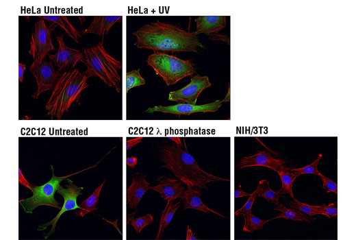
Confocal immunofluorescent analysis of HeLa cells, untreated (top left) or UV-treated (top right), C2C12 cells, untreated (bottom left) or treated with λ phosphatase (bottom middle), and NIH/3T3 cells (bottom right) using Phospho-HSP27 (Ser82) (D1H2F6) XP® Rabbit mAb (Alexa Fluor® 488 Conjugate) (green). Actin filaments were labeled with DY-554 phalloidin (red). Blue pseudocolor = DRAQ5® #4084 (fluorescent DNA dye). Negative staining in NIH/3T3 cells is in agreement with the observation that NIH/3T3 cells do not express HSP27 under basal conditions (5,7).
Go to product page
Image may be subject to copyright.