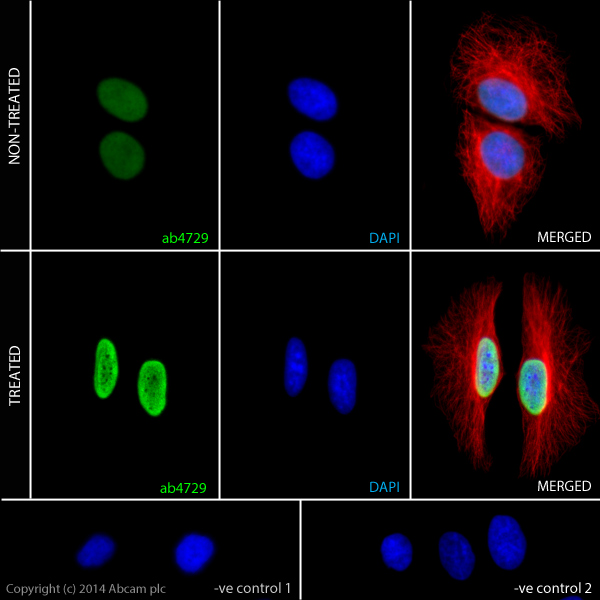
ab4729 staining Histone H3 (acetyl K27) in HeLa cells. The cells were incubated with 10mM Sodium butyrate (ab120948) for 6 hours (Treated) or solvent-only for control purposes (Non-treated). Cells were fixed with 100% methanol (5min) and then blocked in 1% BSA/10% normal goat serum/0.3M glycine in 0.1%PBS-Tween for 1h. The cells were then incubated with ab4729 at 0.5µg/ml and ab7291 at 1µg/ml overnight at +4°C, followed by a further incubation at room temperature for 1h with a anti-rabbit AlexaFluor®488 secondary antibody (ab150077) at 2 µg/ml (shown in green) and a goat anti-mouse AlexaFluor®594 (ab150120) at 2 µg/ml (shown in pseudo colour red). Nuclear DNA was labelled in blue with DAPI. Negative controls: 1– Rabbit primary and anti-mouse secondary antibody; 2 – Mouse primary antibody and anti-rabbit secondary antibody. Controls 1 and 2 indicate that there is no unspecific reaction between primary and secondary antibodies used.
Go to product page
Image may be subject to copyright.