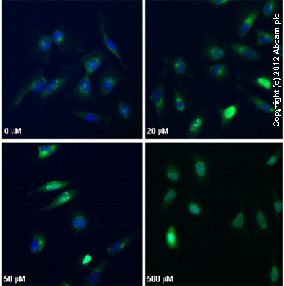
ab2893 staining γH2AX (phospho S139) in HeLa cells treated with camptothecin (ab120115), by ICC/IF. Increased nuclear expression of γH2AX (phospho S139) correlates with increased concentration of camptothecin, as described in literature.The cells were incubated at 37°C for 3h in media containing different concentrations of ab120115 (camptothecin) in DMSO, fixed with 4% formaldehyde for 10 minutes at room temperature and blocked with PBS containing 10% goat serum, 0.3 M glycine, 1% BSA and 0.1% tween for 2h at room temperature. Staining of the treated cells with ab2893 (10 µg/ml) was performed overnight at 4°C in PBS containing 1% BSA and 0.1% tween. A DyLight 488 goat anti-rabbit polyclonal antibody (ab96899) at 1/250 dilution was used as the secondary antibody. Nuclei were counterstained with DAPI and are shown in blue.
Go to product page
Image may be subject to copyright.