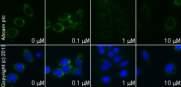
ab110325 staining cytochrome C in MCF7 cells treated with 15-Deoxy-delta12,14-prostaglandin J2 (ab141717), by ICC/IF. Expression of cytochrome C changes from mitochondrial puncta to a difuse staining pattern with increased concentration of 15-Deoxy-delta12,14-prostaglandin J2, as described in literature.The cells were incubated at 37°C for 24 hours in media containing different concentrations of ab141717 (15-Deoxy-delta12,14-prostaglandin J2) in DMSO, fixed with 100% methanol for 5 minutes at -20°C and blocked with PBS containing 10% goat serum, 0.3 M glycine, 1% BSA and 0.1% tween for 2h at room temperature. Staining of the treated cells with ab110325 (10 µg/ml) was performed overnight at 4°C in PBS containing 1% BSA and 0.1% tween. A DyLight 488 anti-mouse polyclonal antibody (ab96879) at 1/250 dilution was used as the secondary antibody. Nuclei (blue) were counterstained with DAPI and membrane is was stained using WGA (red).
Go to product page
Image may be subject to copyright.