Anti-HIF-1-alpha antibody [H1alpha67] - ChIP Grade
| Name | Anti-HIF-1-alpha antibody [H1alpha67] - ChIP Grade |
|---|---|
| Supplier | Abcam |
| Catalog | ab1 |
| Prices | $403.00 |
| Sizes | 100 µg |
| Host | Mouse |
| Clonality | Monoclonal |
| Isotype | IgG2b |
| Clone | H1alpha67 |
| Applications | IHC-P IP ChIP IHC-F FC ICC/IF ICC/IF WB |
| Species Reactivities | Human, Mouse, Rat, Sheep, Rabbit, Bovine, Pig, Ferret, Monkey |
| Antigen | Fusion protein corresponding to Human HIF-1-alpha aa 400-550 |
| Description | Mouse Monoclonal |
| Gene | HIF1A |
| Conjugate | Unconjugated |
| Supplier Page | Shop |
Product images
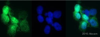 ab1 at 1/200 dilution staining HIF-1-alpha in human 293FT cells by Immunocytochemistry/ Immunofluorescence. Cells were fixed in formaldehyde, permeabilized in 0.5% Trition X-100 and blocked in 5% BSA for 1 hour at 25°C. The primary antibody was used at 1/200 dilution in PBS and incubated with sample at 4°C for 12 hours. An Alexa Fluor® 488 conjugated Goat polyclonal to mouse IgG was used as secondary at 1/500 dilution.See Abreview
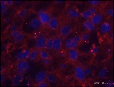 ab1 staining HIF-1-alpha in mouse liver tissue section by Immunohistochemistry (Frozen sections). Tissue samples were fixed with formaldehyde and permeablized with 0.2% Triton-X100 before blocking with 2% BSA for 30 minutes at 20°C. The sample was incubated with primary antibody (1/200) for 9 hours at 4°C. An Alexa Fluor®555-conjugated Goat polyclonal to mouse IgG was used as secondary antibody at 1/200 dilution. DAPI was used to stain the cell nuclei (blue).See Abreview
![All lanes : Anti-HIF-1-alpha antibody [H1alpha67] - ChIP Grade (ab1) at 5 µg/mlLane 1 : HeLa (Human epithelial carcinoma cell line) Nuclear Lysate (ab150036)Lane 2 : Hela-DFO treated (0.5mM, 24h) Nuclear Lysate (ab180880)Lysates/proteins at 40 µg per lane.SecondaryGoat Anti-Mouse IgG H&L (HRP) preadsorbed (ab97040) at 1/10000 dilutiondeveloped using the ECL techniquePerformed under reducing conditions.](http://www.bioprodhub.com/system/product_images/ab_products/2/sub_3/573_ab1-210524-WBAP15789532.jpg) All lanes : Anti-HIF-1-alpha antibody [H1alpha67] - ChIP Grade (ab1) at 5 µg/mlLane 1 : HeLa (Human epithelial carcinoma cell line) Nuclear Lysate (ab150036)Lane 2 : Hela-DFO treated (0.5mM, 24h) Nuclear Lysate (ab180880)Lysates/proteins at 40 µg per lane.SecondaryGoat Anti-Mouse IgG H&L (HRP) preadsorbed (ab97040) at 1/10000 dilutiondeveloped using the ECL techniquePerformed under reducing conditions.
All lanes : Anti-HIF-1-alpha antibody [H1alpha67] - ChIP Grade (ab1) at 5 µg/mlLane 1 : HeLa (Human epithelial carcinoma cell line) Nuclear Lysate (ab150036)Lane 2 : Hela-DFO treated (0.5mM, 24h) Nuclear Lysate (ab180880)Lysates/proteins at 40 µg per lane.SecondaryGoat Anti-Mouse IgG H&L (HRP) preadsorbed (ab97040) at 1/10000 dilutiondeveloped using the ECL techniquePerformed under reducing conditions.
![All lanes : Anti-HIF-1-alpha antibody [H1alpha67] - ChIP Grade (ab1) at 5 µg/mlLane 1 : Hela-Vehicle treated (Negative Control) Whole Cell Lysate (ab116321)Lane 2 : Hela-DFO treated (0.5mM, 24h) Whole Cell Lysate (ab116322)Lysates/proteins at 25 µg per lane.SecondaryGoat Anti-Mouse IgG H&L (HRP) preadsorbed (ab97040) at 1/5000 dilutiondeveloped using the ECL techniquePerformed under reducing conditions.](http://www.bioprodhub.com/system/product_images/ab_products/2/sub_3/574_HIF1-alpha-Primary-antibodies-ab1-24.jpg) All lanes : Anti-HIF-1-alpha antibody [H1alpha67] - ChIP Grade (ab1) at 5 µg/mlLane 1 : Hela-Vehicle treated (Negative Control) Whole Cell Lysate (ab116321)Lane 2 : Hela-DFO treated (0.5mM, 24h) Whole Cell Lysate (ab116322)Lysates/proteins at 25 µg per lane.SecondaryGoat Anti-Mouse IgG H&L (HRP) preadsorbed (ab97040) at 1/5000 dilutiondeveloped using the ECL techniquePerformed under reducing conditions.
All lanes : Anti-HIF-1-alpha antibody [H1alpha67] - ChIP Grade (ab1) at 5 µg/mlLane 1 : Hela-Vehicle treated (Negative Control) Whole Cell Lysate (ab116321)Lane 2 : Hela-DFO treated (0.5mM, 24h) Whole Cell Lysate (ab116322)Lysates/proteins at 25 µg per lane.SecondaryGoat Anti-Mouse IgG H&L (HRP) preadsorbed (ab97040) at 1/5000 dilutiondeveloped using the ECL techniquePerformed under reducing conditions.
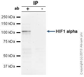 HIF-1-alpha was immunoprecipitated using 0.5mg HeLa Nuclear DFO treated whole cell extract (ab180880), 5µg of Mouse monoclonal to HIF-1-alpha and 50µl of protein G magnetic beads (+). No antibody was added to the control (-).The antibody was incubated under agitation with Protein G beads for 10min, HeLa DFO treated whole cell extract lysate diluted in RIPA buffer was added to each sample and incubated for a further 10min under agitation.Proteins were eluted by addition of 40µl SDS loading buffer and incubated for 10min at 70°C; 10µl of each sample was separated on a SDS PAGE gel, transferred to a nitrocellulose membrane, blocked with 5% BSA and probed with ab1.Secondary: Goat polyclonal to mouse IgG light chain specific (HRP) at 1:20,000 dilution.Band: 110kDa; HIF1 alpha
![Anti-HIF-1-alpha antibody [H1alpha67] - ChIP Grade (ab1) at 1/400 dilution + Human Cell lysate - whole cell (human lung adenocarcinoma cell line ADLC-5M2) treated for 16 hours with 100 micromolar deferoxamine (DFO) at 20 µgPerformed under reducing conditions.](http://www.bioprodhub.com/system/product_images/ab_products/2/sub_3/576_ab1_2.jpg) Anti-HIF-1-alpha antibody [H1alpha67] - ChIP Grade (ab1) at 1/400 dilution + Human Cell lysate - whole cell (human lung adenocarcinoma cell line ADLC-5M2) treated for 16 hours with 100 micromolar deferoxamine (DFO) at 20 µgPerformed under reducing conditions.
Anti-HIF-1-alpha antibody [H1alpha67] - ChIP Grade (ab1) at 1/400 dilution + Human Cell lysate - whole cell (human lung adenocarcinoma cell line ADLC-5M2) treated for 16 hours with 100 micromolar deferoxamine (DFO) at 20 µgPerformed under reducing conditions.
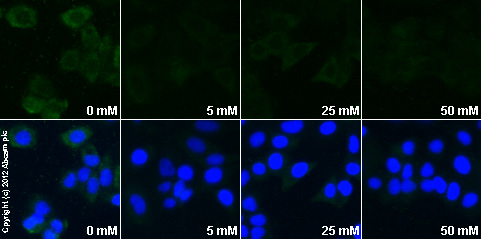 ab1 staining HIF-1-alpha in MCF7 cells treated with metformin hydrochloride (ab120847), by ICC/IF. Decrease in HIF-1-alpha expression correlates with increased concentration of metformin hydrochloride, as described in literature.The cells were incubated at 37°C for 24h in media containing different concentrations of ab120847 (metformin hydrochloride) in water, fixed with 4% formaldehyde for 10 minutes at room temperature and blocked with PBS containing 10% goat serum, 0.3 M glycine, 1% BSA and 0.1% tween for 2h at room temperature. Staining of the treated cells with ab1 (10 µg/ml) was performed overnight at 4°C in PBS containing 1% BSA and 0.1% tween. A goat anti-mouse DyLight 488 antibody (ab96879) at 1/250 dilution was used as the secondary antibody. Nuclei were counterstained with DAPI and are shown in blue.
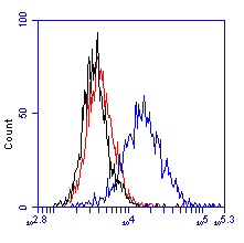 Flow cytometry using ab1. HeLa cells were cultured untreated or with 1mM Deferoxamine (ab120727) for 24 hours to induce HIF-1-alpha protein levels. Cells were then trypsinized, fixed with paraformaldehyde and stained with ab1 (0.5 micrograms/mL). 1% BSA in PBS was used as the blocking buffer throughout. ab1 was labeled with and anti-mouse Alexa-488 dye. Unstained (black), untreated (red) and DFO treated (blue) cell traces are shown.
Product References
HIF-mediated metabolic switching in bladder outlet obstruction mitigates the - HIF-mediated metabolic switching in bladder outlet obstruction mitigates the
Ekman M, Uvelius B, Albinsson S, Sward K. Lab Invest. 2014 May;94(5):557-68.
Conversion of differentiated cancer cells into cancer stem-like cells in a - Conversion of differentiated cancer cells into cancer stem-like cells in a
Auffinger B, Tobias AL, Han Y, Lee G, Guo D, Dey M, Lesniak MS, Ahmed AU. Cell Death Differ. 2014 Jul;21(7):1119-31.
Nanodrug-enhanced radiofrequency tumor ablation: effect of micellar or liposomal - Nanodrug-enhanced radiofrequency tumor ablation: effect of micellar or liposomal
Moussa M, Goldberg SN, Kumar G, Sawant RR, Levchenko T, Torchilin VP, Ahmed M. PLoS One. 2014 Aug 18;9(8):e102727.
Contrasting hypoxic effects on breast cancer stem cell hierarchy is dependent on - Contrasting hypoxic effects on breast cancer stem cell hierarchy is dependent on
Harrison H, Rogerson L, Gregson HJ, Brennan KR, Clarke RB, Landberg G. Cancer Res. 2013 Feb 15;73(4):1420-33.
Thrombin, a mediator of cerebrovascular inflammation in AD and hypoxia. - Thrombin, a mediator of cerebrovascular inflammation in AD and hypoxia.
Tripathy D, Sanchez A, Yin X, Luo J, Martinez J, Grammas P. Front Aging Neurosci. 2013 May 9;5:19.
High glucose enhances HIV entry into T cells through upregulation of CXCR4. - High glucose enhances HIV entry into T cells through upregulation of CXCR4.
Lan X, Cheng K, Chandel N, Lederman R, Jhaveri A, Husain M, Malhotra A, Singhal PC. J Leukoc Biol. 2013 Oct;94(4):769-77.
Combined targeting of PDK1 and EGFR triggers regression of glioblastoma by - Combined targeting of PDK1 and EGFR triggers regression of glioblastoma by
Velpula KK, Bhasin A, Asuthkar S, Tsung AJ. Cancer Res. 2013 Dec 15;73(24):7277-89.
HIV "elite controllers" are characterized by a high frequency of memory CD8+ - HIV "elite controllers" are characterized by a high frequency of memory CD8+
Carriere M, Lacabaratz C, Kok A, Benne C, Jenabian MA, Casartelli N, Hue S, Hocqueloux L, Lelievre JD, Levy Y. J Infect Dis. 2014 May 1;209(9):1321-30.
Glucose-induced O(2) consumption activates hypoxia inducible factors 1 and 2 in - Glucose-induced O(2) consumption activates hypoxia inducible factors 1 and 2 in
Bensellam M, Duvillie B, Rybachuk G, Laybutt DR, Magnan C, Guiot Y, Pouyssegur J, Jonas JC. PLoS One. 2012;7(1):e29807.
Interaction with ErbB4 promotes hypoxia-inducible factor-1alpha signaling. - Interaction with ErbB4 promotes hypoxia-inducible factor-1alpha signaling.
Paatero I, Jokilammi A, Heikkinen PT, Iljin K, Kallioniemi OP, Jones FE, Jaakkola PM, Elenius K. J Biol Chem. 2012 Mar 23;287(13):9659-71.