Anti-HIF-1-alpha antibody [mgc3]
| Name | Anti-HIF-1-alpha antibody [mgc3] |
|---|---|
| Supplier | Abcam |
| Catalog | ab16066 |
| Prices | $404.00 |
| Sizes | 100 µl |
| Host | Mouse |
| Clonality | Monoclonal |
| Isotype | IgG1 |
| Clone | mgc3 |
| Applications | FC Immunoelectrophoresis EMSA EMSA ICC/IF ICC/IF IP WB IHC-P |
| Species Reactivities | Mouse, Bovine, Human, Pig, Primate |
| Antigen | Recombinant fragment corresponding to Human HIF-1-alpha aa 530-826 (C terminal) |
| Description | Mouse Monoclonal |
| Gene | HIF1A |
| Conjugate | Unconjugated |
| Supplier Page | Shop |
Product images
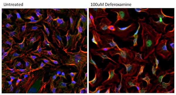 Immunocytochemistry/Immunofluorescent analysis of HIF-1-alpha (green) in HeLa cells either left untreated (left panel) or treated with 100uM Deferoxamine mesylate for ~16 hours (right panel). Formalin-fixed cells were permeabilized with 0.1% Triton X-100 in TBS for 15 minutes at room temperature and blocked with 0.3% BSA for 15 minutes at room temperature. Cells were probed with ab16066 at a dilution of 1/100 for at least 1 hour at room temperature and washed with PBS. Cells were then incubated with a DyLight 488-conjugated goat anti-mouse IgG secondary antibody at a dilution of 1/500 for 30 minutes at room temperature. F-actin (red) was stained with DyLight 594 Phalloidin and nuclei (blue) were stained with Hoechst dye.
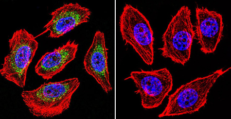 Immunocytochemistry/Immunofluorescence analysis of U251 cells labeling HIF-1-alpha with ab16066. HIF-1-alpha (green), F-Actin staining with Phalloidin (red) and nuclei with DAPI (blue) is shown. Cells were grown on chamber slides and fixed with formaldehyde prior to staining. Cells were probed without (control - right) or with ab16066 (left) at a dilution of 1/20 over night at 4°C and incubated with a DyLight-488 conjugated secondary antibody. Images were taken at 60X magnification.
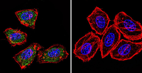 Immunocytochemistry/Immunofluorescence analysis of HeLa cells labeling HIF-1-alpha with ab16066. HIF-1 alpha (green), F-Actin staining with Phalloidin (red) and nuclei with DAPI (blue) is shown. Cells were grown on chamber slides and fixed with formaldehyde prior to staining. Cells were probed without (control - right) or with ab16066 (left) at a dilution of 1/20 over night at 4°C and incubated with a DyLight-488 conjugated secondary antibody. Images were taken at 60X magnification.
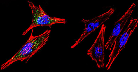 Immunocytochemistry/Immunofluorescence analysis of A2058 cells labeling HIF-1-alpha with ab16066. HIF-1-alpha (green), F-Actin staining with Phalloidin (red) and nuclei with DAPI (blue) is shown. Cells were grown on chamber slides and fixed with formaldehyde prior to staining. Cells were probed without (control - right) or with ab16066 (left) at a dilution of 1/20 over night at 4°C and incubated with a DyLight-488 conjugated secondary antibody. Images were taken at 60X magnification.
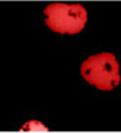
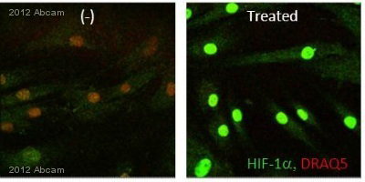 Immunocytochemistry/Immunofluorescence analysis of Human fibroblasts labeling HIF-1-alpha (green) with ab16066.Cells were fixed with paraformaldehyde, permeabilized with 0.3% Triton X-100 and blocked with 5% serum for 1 hour at 24°C. Samples were incubated with primary antibody (1/500 in 0.3% Triton X-100 + 1% BSA) for 1 hour 30 minutes at 24°C. An AlexaFluor®488-conjugated goat anti-mouse polyclonal IgG (1/2000) was used as the secondary antibody.See Abreview
 Immunocytochemistry/Immunofluorescence labeling HIF-1-alpha with ab16066.
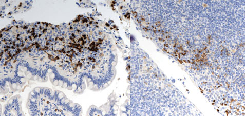 ab16066 staining HIF-1-alpha in Human small intestine (IBD) and tonsil tissue sections by Immunohistochemistry (IHC-P - paraformaldehyde-fixed, paraffin-embedded sections). Tissue was fixed with formaldehyde; antigen retrieval was by heat mediation with an EDTA buffer (pH 9.0). Samples were incubated with primary antibody (1/800 in diluent + background reducers) for 20 minutess at 25°C. An undiluted Goat polymer was used as the secondary antibody.See Abreview
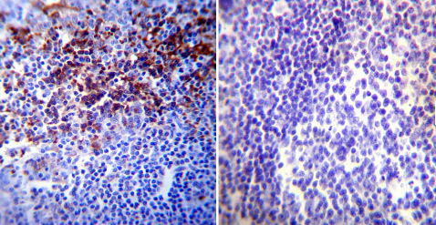 Immunohistochemistry was performed on normal biopsies of deparaffinized Human tonsil tissue. To expose target proteins heat induced antigen retrieval was performed using 10mM sodium citrate (pH6.0) buffer microwaved for 8-15 minutes. Following antigen retrieval tissues were blocked in 3% BSA-PBS for 30 minutes at room temperature. Tissues were then probed at a dilution of 1/20 with ab16066 (left) or without primary antibody (negative control - right) overnight at 4°C in a humidified chamber. Tissues were washed extensively with PBST and endogenous peroxidase activity was quenched with a peroxidase suppressor. Detection was performed using a biotin-conjugated secondary antibody and SA-HRP followed by colorimetric detection using DAB. Tissues were counterstained with hematoxylin and prepped for mounting.
![Anti-HIF-1-alpha antibody [mgc3] (ab16066) at 1/2000 dilution + HeLa cell lysate](http://www.bioprodhub.com/system/product_images/ab_products/2/sub_3/593_ab16066_1.jpg) Anti-HIF-1-alpha antibody [mgc3] (ab16066) at 1/2000 dilution + HeLa cell lysate
Anti-HIF-1-alpha antibody [mgc3] (ab16066) at 1/2000 dilution + HeLa cell lysate
![Overlay histogram showing HeLa cells stained with ab16066 (red line). The cells were fixed with 80% methanol (5 min) and then permeabilized with 0.1% PBS-Tween for 20 min. The cells were then incubated in 1x PBS / 10% normal goat serum / 0.3M glycine to block non-specific protein-protein interactions followed by the antibody (ab16066, 2µg/1x106 cells) for 30 min at 22ºC. The secondary antibody used was DyLight® 488 goat anti-mouse IgG (H+L) (ab96879) at 1/500 dilution for 30 min at 22ºC. Isotype control antibody (black line) was mouse IgG1 [ICIGG1] (ab91353, 2µg/1x106 cells) used under the same conditions. Acquisition of >5,000 events was performed.](http://www.bioprodhub.com/system/product_images/ab_products/2/sub_3/594_HIF1-alpha-Primary-antibodies-ab16066-10.jpg) Overlay histogram showing HeLa cells stained with ab16066 (red line). The cells were fixed with 80% methanol (5 min) and then permeabilized with 0.1% PBS-Tween for 20 min. The cells were then incubated in 1x PBS / 10% normal goat serum / 0.3M glycine to block non-specific protein-protein interactions followed by the antibody (ab16066, 2µg/1x106 cells) for 30 min at 22ºC. The secondary antibody used was DyLight® 488 goat anti-mouse IgG (H+L) (ab96879) at 1/500 dilution for 30 min at 22ºC. Isotype control antibody (black line) was mouse IgG1 [ICIGG1] (ab91353, 2µg/1x106 cells) used under the same conditions. Acquisition of >5,000 events was performed.
Overlay histogram showing HeLa cells stained with ab16066 (red line). The cells were fixed with 80% methanol (5 min) and then permeabilized with 0.1% PBS-Tween for 20 min. The cells were then incubated in 1x PBS / 10% normal goat serum / 0.3M glycine to block non-specific protein-protein interactions followed by the antibody (ab16066, 2µg/1x106 cells) for 30 min at 22ºC. The secondary antibody used was DyLight® 488 goat anti-mouse IgG (H+L) (ab96879) at 1/500 dilution for 30 min at 22ºC. Isotype control antibody (black line) was mouse IgG1 [ICIGG1] (ab91353, 2µg/1x106 cells) used under the same conditions. Acquisition of >5,000 events was performed.
Product References
PINK1 deficiency sustains cell proliferation by reprogramming glucose metabolism - PINK1 deficiency sustains cell proliferation by reprogramming glucose metabolism
Requejo-Aguilar R, Lopez-Fabuel I, Fernandez E, Martins LM, Almeida A, Bolanos JP. Nat Commun. 2014 Jul 24;5:4514.
Autophagy promotes paclitaxel resistance of cervical cancer cells: involvement of - Autophagy promotes paclitaxel resistance of cervical cancer cells: involvement of
Peng X, Gong F, Chen Y, Jiang Y, Liu J, Yu M, Zhang S, Wang M, Xiao G, Liao H. Cell Death Dis. 2014 Aug 14;5:e1367.
Fusicoccin a, a phytotoxic carbotricyclic diterpene glucoside of fungal origin, - Fusicoccin a, a phytotoxic carbotricyclic diterpene glucoside of fungal origin,
Bury M, Andolfi A, Rogister B, Cimmino A, Megalizzi V, Mathieu V, Feron O, Evidente A, Kiss R. Transl Oncol. 2013 Apr;6(2):112-23. Epub 2013 Apr 1.
Melatonin: the watchdog of villous trophoblast homeostasis against - Melatonin: the watchdog of villous trophoblast homeostasis against
Lanoix D, Lacasse AA, Reiter RJ, Vaillancourt C. Mol Cell Endocrinol. 2013 Dec 5;381(1-2):35-45.
Prognostic impact of PHIP copy number in melanoma: linkage to ulceration. - Prognostic impact of PHIP copy number in melanoma: linkage to ulceration.
Bezrookove V, De Semir D, Nosrati M, Tong S, Wu C, Thummala S, Dar AA, Leong SP, Cleaver JE, Sagebiel RW, Miller JR 3rd, Kashani-Sabet M. J Invest Dermatol. 2014 Mar;134(3):783-90.
Fibulin-5 is up-regulated by hypoxia in endothelial cells through a - Fibulin-5 is up-regulated by hypoxia in endothelial cells through a
Guadall A, Orriols M, Rodriguez-Calvo R, Calvayrac O, Crespo J, Aledo R, Martinez-Gonzalez J, Rodriguez C. J Biol Chem. 2011 Mar 4;286(9):7093-103.
Endogenous myoglobin in human breast cancer is a hallmark of luminal cancer - Endogenous myoglobin in human breast cancer is a hallmark of luminal cancer
Kristiansen G, Rose M, Geisler C, Fritzsche FR, Gerhardt J, Luke C, Ladhoff AM, Knuchel R, Dietel M, Moch H, Varga Z, Theurillat JP, Gorr TA, Dahl E. Br J Cancer. 2010 Jun 8;102(12):1736-45.
SDHB loss predicts malignancy in pheochromocytomas/sympathethic paragangliomas, - SDHB loss predicts malignancy in pheochromocytomas/sympathethic paragangliomas,
Blank A, Schmitt AM, Korpershoek E, van Nederveen F, Rudolph T, Weber N, Strebel RT, de Krijger R, Komminoth P, Perren A. Endocr Relat Cancer. 2010 Oct 5;17(4):919-28.
Physiologic oxygen concentration enhances the stem-like properties of CD133+ - Physiologic oxygen concentration enhances the stem-like properties of CD133+
McCord AM, Jamal M, Shankavaram UT, Lang FF, Camphausen K, Tofilon PJ. Mol Cancer Res. 2009 Apr;7(4):489-97.
Loss of VHL and hypoxia provokes PAX2 up-regulation in clear cell renal cell - Loss of VHL and hypoxia provokes PAX2 up-regulation in clear cell renal cell
Luu VD, Boysen G, Struckmann K, Casagrande S, von Teichman A, Wild PJ, Sulser T, Schraml P, Moch H. Clin Cancer Res. 2009 May 15;15(10):3297-304.