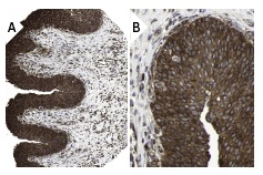
NFκB p65 (F-6): sc-8008. Immunoperoxidase staining of formalin fixed, paraffin-embedded human urinary bladder tissue showing cytoplasmic staining of surface epithelial cells at low (A) and high (B) magnification. Kindly provided by The Swedish Human Protein Atlas (HPA) program.
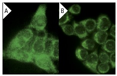
NFκB p65 (F-6): sc-8008. Immunofluorescence staining of methanol-fixed A-431 cells showing cytoplasmic localization using indirect FITC (A) staining and HeLa cells using direct Alexa Fluor 488 (B) staining.
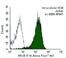
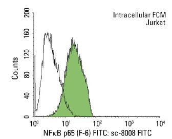
NFκB p65 (F-6) Alexa Fluor 647: sc-8008 AF647. Intracellular FCM analysis of fixed and permeabilized Jurkat cells. Black line histogram represents the isotype control, normal mouse IgG
1: sc-24636.
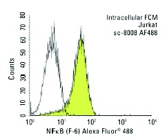
NFκB p65 (F-6) Alexa Fluor 488: sc-8008 AF488. Intracellular FCM analysis of fixed and permeabilized Jurkat cells. Black line histogram represents the isotype control, normal mouse IgG
1: sc-3890.
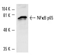
NFκB p65 (F-6): sc-8008. Western blot analysis of NFκB p65 expression in Jurkat whole cell lysate.
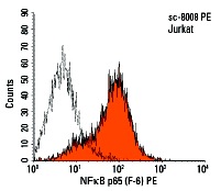
NFκB p65 (F-6) PE: sc-8008 PE. Intracellular FCM analysis of fixed and permeabilized Jurkat cells. Black line histogram represents the isotype control, normal mouse IgG
1: sc-2866.
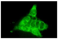
NFκB p65 (F-6): sc-8008. Immunofluorescence staining of methanol-fixed A-431 cells showing cytoplasmic localization.
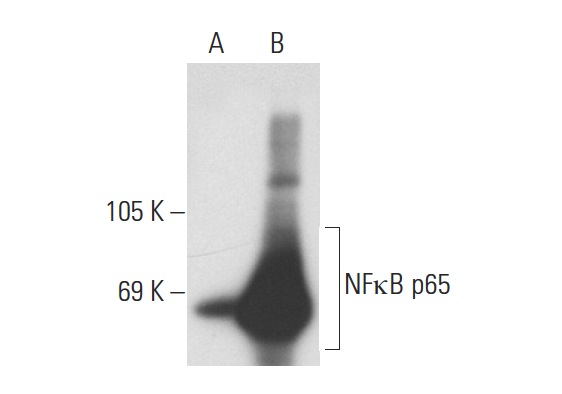
NFκB p65 (F-6): sc-8008. Western blot analysis of NFκB p65 expression in non-transfected: sc-117752 (A) and mouse NFκB p65 transfected: sc-122028 (B) 293T whole cell lysates.
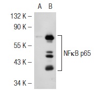
NFκB p65 (F-6): sc-8008. Western blot analysis of NFκB p65 expression in non-transfected: sc-117752 (A) and mouse NFκB p65 transfected: sc-122027 (B) 293T whole cell lysates.
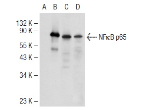
NFκB p65 (F-6): sc-8008. Western blot analysis of NFκB p65 expression in non-transfected 293T: sc-117752 (A), mouse NFκB p65 transfected 293T: sc-122028 (B), HUV-EC-C (C) and NIH/3T3 (D) whole cell lysates.
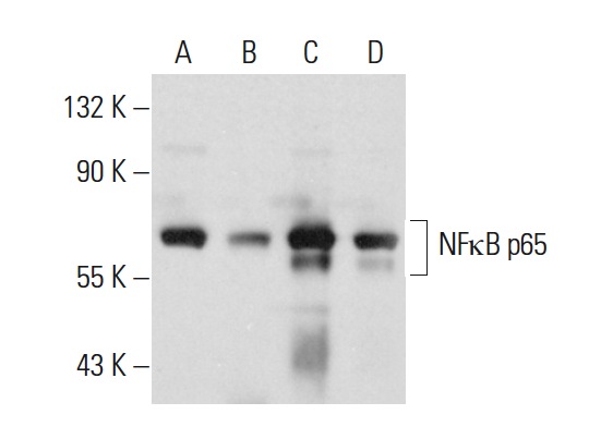
NFκB p65 (F-6): sc-8008. Western blot analysis of NFκB p65 expression in HeLa (A), MIA PaCa-2 (B), T24 (C) and SK-BR-3 (D) whole cell lysates.
NFκB p65 (F-6) FITC: sc-8008 FITC. Intracellular FCM analysis of fixed and permeabilized Jurkat cells. Black line histogram represents the isotype control, normal mouse IgG
1: sc-2855.
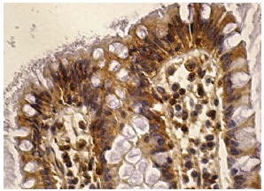
NFκB p65 (F-6): sc-8008. Immunoperoxidase staining of formalin fixed, paraffin-embedded human colon tissue showing cytoplasmic staining of glandular cells.