Anti-TRPM1 Antibody, clone 545H5
| Name | Anti-TRPM1 Antibody, clone 545H5 |
|---|---|
| Supplier | Sigma-Aldrich |
| Catalog | MABN1841 |
| Prices | $299.00 |
| Sizes | mabn1841 |
| Clonality | Monoclonal |
| Isotype | IgG2bκ |
| Applications | WB |
| Species Reactivities | Mouse |
| Purity/Format | Protein G Purified |
| Description | Monoclonal |
| Gene | Trpm1 |
| Supplier Page | Shop |
Product images
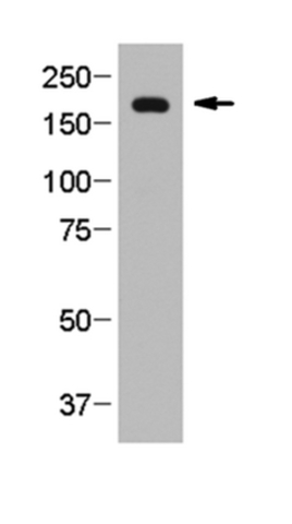 Western Blotting Analysis:
Representative lot data.
Proteins in mouse retina post-nuclear lysate were seperated by electrophoresis (~25 µg or ~0.3 retina equivalent per lane) without prior heating, transferred to nitrocellulose membrane, blocked with 5% milk in TBST, and probed with Cat. No. MABN1841, Anti-TRPM1, clone 545H5 (1 µg/mL) overnight at 4°C. Proteins were visualized using an anti-mouse IgG secondary antibody conjugated to HRP and a chemiluminescence detection system.
Arrow indicates TRPM1 (~180 kDa).
(Image courtesy of Dr. Melina A. Agosto, Baylor College of Medicine, Houston, TX)