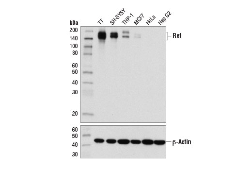
Western blot analysis of extracts from various cell lines using Ret (E1N8X) XP ® Rabbit mAb (upper) and β-Actin (D6A8) Rabbit mAb #8457 (lower).

Immunohistochemical analysis of paraffin-embedded human adrenal gland using Ret (E1N8X) XP ® Rabbit mAb.

Immunohistochemical analysis of paraffin-embedded human breast carcinoma using Ret (E1N8X) XP ® Rabbit mAb.

Immunohistochemical analysis of paraffin-embedded human small intestine using Ret (E1N8X) XP ® Rabbit mAb. Note the positive signal of the myenteric plexus in the muscularis externa of the small intestine.

Immunohistochemical analysis of paraffin-embedded TT (left) or Hep G2 (right) cell pellets using Ret (E1N8X) XP ® Rabbit mAb.

Confocal immunofluorescent analysis of TT (positive, left) and HeLa (negative, right) cells using Ret (E1N8X) XP ® Rabbit mAb (green). Blue pseudocolor = DRAQ5 ® #4084 (fluorescent DNA dye).

Flow cytometric analysis of TT cells (positive, green) and HeLa cells (negative, blue) using Ret (E1N8X) XP ® Rabbit mAb. Anti-rabbit IgG (H+L), F(ab') 2 Fragment (Alexa Fluor ® 488 Conjugate) #4412 was used as a secondary antibody.