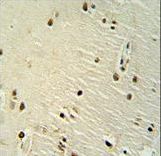
XRCC6 antibody immunohistochemistry of formalin-fixed and paraffin-embedded human brain tissue followed by peroxidase-conjugated secondary antibody and DAB staining.
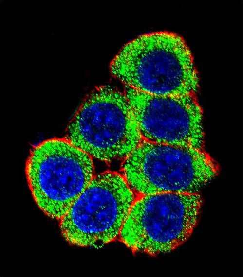
Confocal immunofluorescent of XRCC6 Antibody with 293 cell followed by Alexa Fluor 488-conjugated goat anti-rabbit lgG (green). Actin filaments have been labeled with Alexa Fluor 555 phalloidin (red). DAPI was used to stain the cell nuclear (blue).
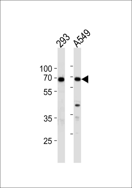
XRCC6 Antibody western blot of 293,A549 cell line lysates (35 ug/lane). The XRCC6 antibody detected the XRCC6 protein (arrow).
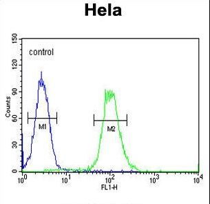
XRCC6 Antibody flow cytometry of HeLa cells (right histogram) compared to a negative control cell (left histogram). FITC-conjugated goat-anti-rabbit secondary antibodies were used for the analysis.