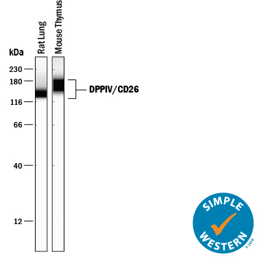
Simple Western lane view shows lysates of rat lung tissue and mouse thymus tissue, loaded at 0.2 mg/mL. A specific band was detected for DPPIV/CD26 at approximately 144-169 kDa (as indicated) using 5 µg/mL of Goat Anti-Mouse DPPIV/CD26 Antigen Affinity-purified Polyclonal Antibody (Catalog # AF954) followed by 1:50 dilution of HRP-conjugated Anti-Goat IgG Secondary Antibody (Catalog # HAF109). This experiment was conducted under reducing conditions and using the 12-230 kDa separation system.
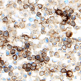
DPPIV/CD26 was detected in immersion fixed frozen sections of mouse thymus using Goat Anti-Mouse DPPIV/CD26 Antigen Affinity-purified Polyclonal Antibody (Catalog # AF954) at 1.7 µg/mL overnight at 4 °C. Tissue was stained using the Anti-Goat HRP-DAB Cell & Tissue Staining Kit (brown; Catalog # CTS008) and counterstained with hematoxylin (blue). Specific staining was localized to lymphocytes. View our protocol for Chromogenic IHC Staining of Frozen Tissue Sections.
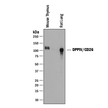
Western blot shows lysates of mouse thymus tissue and rat lung tissue. PVDF membrane was probed with 0.25 µg/mL of Goat Anti-Mouse DPPIV/CD26 Antigen Affinity-purified Polyclonal Antibody (Catalog # AF954) followed by HRP-conjugated Anti-Goat IgG Secondary Antibody (Catalog # HAF019). A specific band was detected for DPPIV/CD26 at approximately 100-110 kDa (as indicated). This experiment was conducted under reducing conditions and using Immunoblot Buffer Group 1.