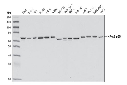
Western blot analysis of extracts from various cell lines using NF-κB p65 (E498) Antibody.

Immunohistochemical analysis of paraffin-embedded human breast carcinoma using NF-κB p65 (E498) Antibody.

Immunohistochemical analysis of paraffin-embedded chronic cholecystitis using NF-κB p65 (E498) Antibody.

Immunohistochemical analysis of paraffin-embedded mouse spleen using NF-κB p65 (E498) Antibody.

Immunohistochemical analysis of paraffin-embedded HeLa cell pellets, untreated (left) or treated with hTNF-α #8902 (right), using NF-κB p65 (E498) Antibody.

Confocal immunofluorescent analysis of HeLa cells, untreated (left) or treated with hTNF-α #8902 (20 ng/ml for 20 minutes) (right), using NF-κB p65 (E498) Antibody (green). Actin filaments were labeled with DY-554 phalloidin (red). Blue pseudocolor = DRAQ5 ® #4084 (fluorescent DNA dye).

Flow cytometric analysis of Jurkat cells using NF-κB p65 (E498) Antibody (blue) compared to Rabbit (DA1E) mAb IgG XP ® Isotype Control #3900 (red).