CREB1

Gene Symbol CREB1
Entrez Gene 281713
Alt Symbol CREB
Species Bovine
Gene Type protein-coding
Description cAMP responsive element binding protein 1
Other Description CREB-1|cAMP-responsive element-binding protein 1|cyclic AMP-responsive DNA-binding protein|cyclic AMP-responsive element-binding protein 1
Swissprots A5PK02 O18957 P27925
Accessions DAA32469 P27925 AF006042 AAB62381 BC142303 AAI42304 BT026534 ABH06321 X57031 CAA40347 NM_174285 NP_776710
Function Phosphorylation-dependent transcription factor that stimulates transcription upon binding to the DNA cAMP response element (CRE), a sequence present in many viral and cellular promoters. Transcription activation is enhanced by the TORC coactivators which act independently of Ser-117 phosphorylation. Involved in different cellular processes including the synchronization of circadian rhythmicity and the differentiation of adipose cells. {ECO:0000269|PubMed:1309910, ECO:0000269|PubMed:8057465, ECO:0000269|PubMed:8627725}.
Subcellular Location Nucleus.
Top Pathways Prostate cancer, Adrenergic signaling in cardiomyocytes, cGMP-PKG signaling pathway, Thyroid hormone synthesis, TNF signaling pathway

Anti-CREB + CREM antibody - ab5803 from Abcam

Prices $370.00
Sizes 100 µg
Host Rabbit
Clonality Polyclonal
Immunofluorescent analysis of CREB (green) showing staining in the nucleus of SK-N-MC cells (right) compared to a negative control without primary antibody (left). Formalin-fixed cells were permeabilized with 0.1% Triton X-100 in TBS for 5-10 minutes and blocked with 3% BSA-PBS for 30 minutes at room temperature. Cells were probed with a CREB polyclonal antibody (ab5803) in 3% BSA-PBS at a dilution of 1:200 and incubated overnight at 4ºC in a humidified chamber. Cells were washed with PBST and incubated with a DyLight-conjugated secondary antibody in PBS at room temperature in the dark. Actin was stained using Alexa Fluor 554 (red) and nuclei were stained with Hoechst or DAPI (blue). Images were taken at a magnification of 60x.

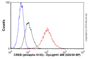
Anti-CREB (phospho S133) antibody [E113] - ab32096 from Abcam

Prices $403.00
Sizes 100 µl
Host Rabbit
Clonality Monoclonal
Clone E113
Overlay histogram showing A431 cells stained with ab32096 (red line). The cells were fixed with 80% methanol (5 min) and then permeabilized with 0.1% PBS-Tween for 20 min. The cells were then incubated in 1x PBS / 10% normal goat serum / 0.3M glycine to block non-specific protein-protein interactions followed by the antibody (ab32096, 1/1000 dilution) for 30 min at 22°C. The secondary antibody used was DyLight® 488 goat anti-rabbit IgG (H+L) (ab96899) at 1/500 dilution for 30 min at 22°C. Isotype control antibody (black line) was rabbit IgG (monoclonal) (0.1μg/1x106 cells) used under the same conditions. Unlabelled sample (blue line) was also used as a control. Acquisition of >5,000 events were collected using a 20mW Argon ion laser (488nm) and 525/30 bandpass filter.

Anti-CREB antibody - ab65741 from Abcam

Prices $376.00
Sizes 100 µg
Host Sheep
Clonality Polyclonal
Anti-CREB antibody (ab65741) at 2 µg/ml + J774.1 (J) cell extracts

Anti-CREB antibody - ab84757 from Abcam

Prices $376.00
Sizes 100 µl
Host Rabbit
Clonality Polyclonal
ab84757, at 1/100 dilution, staining CREB in formalin-fixed, paraffin-embedded human prostate carcinoma by Immunohistochemistry using a Red-fluorescent goat anti-rabbit IgG highly cross-adsorbed Hilyte PlusTM 555 secondary antibody at a dilution of 1/100.

Anti-CREB antibody - ab178539 from Abcam

Prices $376.00
Sizes 200 µl
Host Goat
Clonality Polyclonal
All lanes : Anti-CREB antibody (ab178539) at 0.1 µg/ml (Incubation 1 hour)Lane 1 : Nuclear lysate from Jurkat cellsLane 2 : Nuclear lysate from NIH3T3 cellsLysates/proteins at 35 µg per lane.developed using the ECL technique

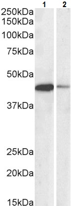
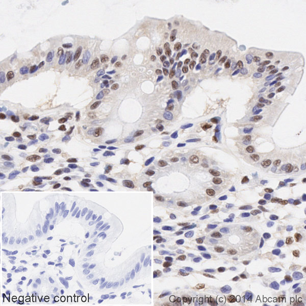
Anti-CREB antibody [E306] - ab32515 from Abcam

Prices $392.00
Sizes 100 µl
Host Rabbit
Clonality Monoclonal
Clone E306
Anti-CREB antibody [E306] (ab32515) at 1/1000 dilution + A431 cell lysate

Anti-CREB antibody [E306] (HRP) - ab194313 from Abcam

Prices $425.00
Sizes 100 µl
Host Rabbit
Clonality Monoclonal
Clone E306
IHC image of CREB staining in a section of formalin-fixed paraffin-embedded normal human colon tissue*, performed on a Leica BOND. The section was pre-treated using heat mediated antigen retrieval with sodium citrate buffer (pH6, epitope retrieval solution 1) for 20mins. The section was then incubated with ab194313 at 1/100 dilution, for 15 mins at room temperature. DAB was used as the chromogen. The section was then counterstained with haematoxylin and mounted with DPX. The inset negative control image is taken from an identical assay without primary antibody.For other IHC staining systems (automated and non-automated) customers should optimize variable parameters such as antigen retrieval conditions, primary antibody concentration and antibody incubation times.*Tissue obtained from the Human Research Tissue Bank, supported by the NIHR Cambridge Biomedical Research Centre.

CREB, Ser133 - MBS602139 from MyBioSource

Clonality Monoclonal
Clone 6k87

CREB, Ser133, Polyclonal Antibody - MBS610102 from MyBioSource

Prices $690.00
Sizes 100 µl
Host Rabbit
Clonality Polyclonal

CREB, Ser133, Polyclonal Antibody - MBS617309 from MyBioSource

Prices $620.00
Sizes 100 µl
Host Rabbit
Clonality Polyclonal

CREB, Ser133, Polyclonal Antibody - MBS611737 from MyBioSource

Prices $630.00
Sizes 100 µl
Host Rabbit
Clonality Polyclonal
Search more