AURKA

Gene Symbol AURKA
Entrez Gene 504437
Alt Symbol ARK-1
Species Bovine
Gene Type protein-coding
Description aurora kinase A
Other Description Aurora-A|aurora 2|aurora-related kinase 1|aurora/IPL1-related kinase 1|serine/threonine protein kinase 6|serine/threonine-protein kinase 15|serine/threonine-protein kinase 6|serine/threonine-protein kinase aurora-A
Swissprots Q2TA06
Accessions DAA23154 Q2TA06 BC111181 AAI11182 DQ334808 ABC61056 XM_005214525 XP_005214582 NM_001038028 NP_001033117
Function Mitotic serine/threonine kinases that contributes to the regulation of cell cycle progression. Associates with the centrosome and the spindle microtubules during mitosis and plays a critical role in various mitotic events including the establishment of mitotic spindle, centrosome duplication, centrosome separation as well as maturation, chromosomal alignment, spindle assembly checkpoint, and cytokinesis. Required for initial activation of CDK1 at centrosomes. Phosphorylates numerous target proteins, including ARHGEF2, BORA, BRCA1, CDC25B, DLGP5, HDAC6, KIF2A, LATS2, NDEL1, PARD3, PPP1R2, PLK1, RASSF1, TACC3, p53/TP53 and TPX2. Regulates KIF2A tubulin depolymerase activity. Required for normal axon formation. Plays a role in microtubule remodeling during neurite extension. Important for microtubule formation and/or stabilization. Also acts as a key regulatory component of the p53/TP53 pathway, and particularly the checkpoint-response pathways critical for oncogenic transformation of cel
Subcellular Location 
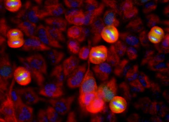
Cytoplasm, cytoskeleton, microtubule organizing center, centrosome {ECO:0000250}. Cytoplasm, cytoskeleton, spindle pole {ECO:0000250}. Note=Detected at the neurite hillock in developing neurons (By similarity). Localizes at the centrosome in mitotic cells from early prophase until telophase, but also localizes to the spindle pole MTs from prophase to anaphase. Moves to the midbody during both telophase and cytokinesis. Associates with both the pericentriolar material (PCM) and centrioles. Colocalized with SIRT2 at centrosome (By similarity). {ECO:0000250}.
Top Pathways Oocyte meiosis

Anti-Aurora A antibody - ab47178 from Abcam

Prices $376.00
Sizes 100 µg
Host Rabbit
Clonality Polyclonal
All lanes : Anti-Aurora A antibody (ab47178) at 2 µg/mlLane 1 : HCT-116 lysateLane 2 : Human testis lysateLane 3 : Mouse testis lysateLane 4 : Rat testis lysate

Anti-Aurora A antibody [1A11] - ab190367 from Abcam

Prices $376.00
Sizes 100 µl
Host Mouse
Clonality Monoclonal
Clone 1A11
Immunofluorescence analysis of HeLa cells, labeling Aurora A using ab190367 at a 1/100 dilution (green). Counterstained with an antibody against Vimentin (red) and DNA (blue).

Aurora A Monoclonal Antibody (35C1) - 45-8900 from Thermo Fisher Scientific

Prices $304.00
Sizes 100 µg
Host Mouse
Clonality Monoclonal
Clone 35C1
Western blot analysis of Aurora A was performed by loading 30 ug of Jurkat (lane1), K562 (lane2) and MCF7 (lane3) cell lysate using Novex® NuPAGE® 4-12 % Bis-Tris gel (NP0321BOX), XCell SureLock™ Electrophoresis System (EI0002), Novex® Sharp Pre-Stained Protein Standard (LC5800), and iBlot® Dry Blotting System (IB21001). Proteins were transferred to a nitrocellulose membrane and blocked with 5 % skim milk for 1 hour at room temperature. Aurora A was detected at ~ 45 kDa using Aurora A Mouse Monoclonal Antibody (458900) at 1-3 ug/ml in 5 % skim milk at 4°C overnight on a rocking platform. Goat Anti-Mouse IgG - HRP Secondary Antibody (626520) 1:4000 dilution was used and chemiluminescent detection was performed using Novex® ECL Chemiluminescent Substrate Reagent Kit (WP20005).
Search more