MAP1LC3A

Gene Symbol MAP1LC3A
Entrez Gene 84557
Alt Symbol ATG8E, LC3, LC3A, MAP1ALC3, MAP1BLC3
Species Human
Gene Type protein-coding
Description microtubule-associated protein 1 light chain 3 alpha
Other Description MAP1 light chain 3-like protein 1|MAP1A/1B light chain 3 A|MAP1A/MAP1B LC3 A|MAP1A/MAP1B light chain 3 A|autophagy-related ubiquitin-like modifier LC3 A|microtubule-associated proteins 1A/1B light chain 3|microtubule-associated proteins 1A/1B light chain 3A
Swissprots Q9BXW5 E1P5P5 E1P5P4 Q9H492
Accessions EAW76263 EAW76264 EAW76265 EAW76266 Q9H492 AF276658 AAK35151 AL833855 CAD38714 AM392590 CAL37468 BC015810 AAH15810 BM740344 BM919877 BP212418 BT007452 AAP36120 DQ893641 ABM84567 DQ895536 ABM86462 EL954540 H18312 XM_011529083 XP_011527385 XM_011529084 XP_011527386 XM_011529085 XP_011527387 NM_032514 NP_115903 NM_181509 NP_852610
Function Ubiquitin-like modifier involved in formation of autophagosomal vacuoles (autophagosomes). Whereas LC3s are involved in elongation of the phagophore membrane, the GABARAP/GATE-16 subfamily is essential for a later stage in autophagosome maturation. {ECO:0000269|PubMed:20713600}.
Subcellular Location Cytoplasm, cytoskeleton. Endomembrane system; Lipid-anchor. Cytoplasmic vesicle, autophagosome membrane; Lipid-anchor. Note=LC3-II binds to the autophagic membranes.
Tissue Specificity Most abundant in heart, brain, liver, skeletal muscle and testis but absent in thymus and peripheral blood leukocytes. {ECO:0000269|PubMed:12740394}.

LC3A/B (D3U4C) XP ® Rabbit mAb (Biotinylated) - 13118 from Cell Signaling Technology

Prices $329.00
Sizes 100 µl (10 western blots)
Host Rabbit
Clonality Monoclonal
Clone D3U4C
Western blot analysis of HeLa cell extracts (LC3 Control Cell Extracts #11972), untreated (-) or chloroquine-treated (50 μM, overnight; +), using LC3A/B (D3U4C) XP ® Rabbit mAb (Biotinylated). Streptavidin-HRP #3999 was used for detection.

LC3A/B (D3U4C) XP ® Rabbit mAb (Alexa Fluor ® 488 Conjugate) - 13082 from Cell Signaling Technology

Prices $329.00
Sizes 100 µl (50 tests)
Host Rabbit
Clonality Monoclonal
Clone D3U4C
Confocal immunofluorescent analysis of HeLa cells, chloroquine-treated (50 μM, overnight; left), nutrient-starved with EBSS (4 hr, middle), or untreated (right) using LC3A/B (D3U4C) XP ® Rabbit mAb (Alexa Fluor ® 488 Conjugate) #13082 (green) and β-Actin (8H10D10) Mouse mAb #3700 (red). Blue pseudocolor = DRAQ5 ® #4084 (fluorescent DNA dye).

LC3A/B (D3U4C) XP ® Rabbit mAb (Pacific Blue™ Conjugate) - 14083 from Cell Signaling Technology

Prices $329.00
Sizes 100 µl (50 tests)
Host Rabbit
Clonality Monoclonal
Clone D3U4C
Flow cytometric analysis of HeLa cells, untreated (blue) or treated with chloroquine (50 μM, 16 hr) (green), using LC3A/B (D3U4C) XP ® Rabbit mAb (Pacific Blue™ Conjugate).

LC3A/B (D3U4C) XP ® Rabbit mAb (Alexa Fluor ® 555 Conjugate) - 13173 from Cell Signaling Technology

Prices $329.00
Sizes 100 µl (50 tests)
Host Rabbit
Clonality Monoclonal
Clone D3U4C
Confocal immunofluorescent analysis of HeLa cells, chloroquine-treated (50 μM, overnight; left), nutrient-starved with EBSS (4 hr, middle), or untreated (right) using LC3A/B (D3U4C) XP ® Rabbit mAb (Alexa Fluor ® 555 Conjugate) #13173 (red) and β-Actin (8H10D10) Mouse mAb #3700 (green). Blue pseudocolor = DRAQ5 ® #4084 (fluorescent DNA dye).

LC3A/B (D3U4C) XP ® Rabbit mAb (Alexa Fluor ® 594 Conjugate) - 14079 from Cell Signaling Technology

Prices $329.00
Sizes 100 µl (50 tests)
Host Rabbit
Clonality Monoclonal
Clone D3U4C
Immunofluorescent analysis of HeLa cells, untreated (left), nutrient-starved with EBSS (3 hr; center), or chloroquine-treated (50 μM, overnight; right), using LC3A/B (D3U4C) XP ® Rabbit mAb (Alexa Fluor ® 594 Conjugate) #14079 (red) and β-Actin (8H10D10) Mouse mAb #3700 (green). Blue pseudocolor= DRAQ5 ® #4084 (fluorescent DNA dye).

LC3A/B (D3U4C) XP ® Rabbit mAb (Alexa Fluor ® 647 Conjugate) - 13394 from Cell Signaling Technology

Prices $329.00
Sizes 100 µl (50 tests)
Host Rabbit
Clonality Monoclonal
Clone D3U4C
Confocal immunofluorescent analysis of HeLa cells, untreated (left), nutrient-starved with EBSS (4 hr; middle), or chloroquine-treated (50 μM, overnight; right), using LC3A/B (D3U4C) XP ® Rabbit mAb (Alexa Fluor ® 647 Conjugate) #13394 (blue) and β-Actin (8H10D10) Mouse mAb #3700 (green). Red = Propidium Iodide (PI)/RNase Staining Solution #4087.

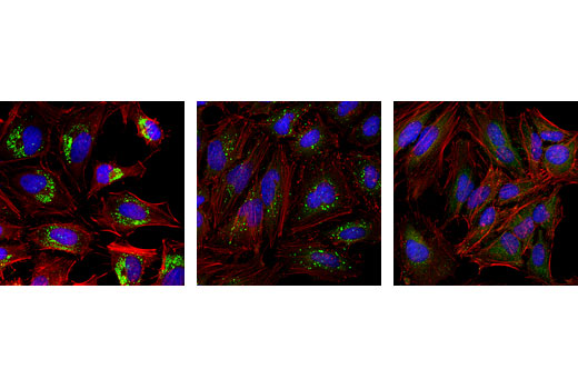
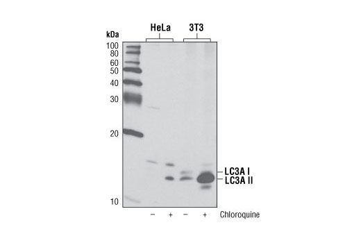
LC3A (D50G8) XP ® Rabbit mAb - 4599 from Cell Signaling Technology

Prices $109.00, $277.00
Sizes 20 µl (2 western blots), 100 µl (10 western blots)
Host Rabbit
Clonality Monoclonal
Clone D50G8
Western blot analysis of extracts from HeLa and NIH/3T3 cells, untreated or chloroquine-treated (50 μM, overnight), using LC3A (D50G8) XP ® Rabbit mAb.

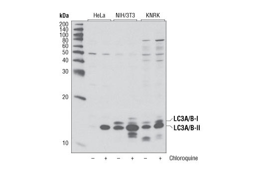
LC3A/B (D3U4C) XP ® Rabbit mAb - 12741 from Cell Signaling Technology

Prices $109.00, $277.00
Sizes 20 µl (2 western blots), 100 µl (10 western blots)
Host Rabbit
Clonality Monoclonal
Clone D3U4C
Western blot analysis of extracts from RD cells, untreated (-) or Torin 1-treated (250 nM, 4 hr; +), using LC3A/B (D3U4C) XP ® Rabbit mAb.

LC3A/B Antibody - 4108 from Cell Signaling Technology

Prices $246.00
Sizes 100 µl (10 western blots)
Host Rabbit
Clonality Polyclonal
Western blot analysis of extracts from various cell lines, untreated or treated with chloroquine (50 μM, overnight) using LC3A/B Antibody.

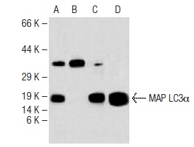
MAP LC3α (R-23) - sc-134226 from Santa Cruz Biotechnology

Prices $295.00
Sizes 100 µg/ml
Host Rabbit
Clonality Polyclonal
MAP LC3α (R-23): sc-134226. Western blot analysis of MAP LC3α expression in human brain tissue extract in the absence (A) and the presence (B) of immunizing peptide, mouse brain (C) tissue extract and rat brain (D) tissue extract.

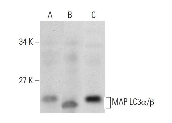
MAP LC3α/β (H-47) - sc-292354 from Santa Cruz Biotechnology

Prices $279.00
Sizes 200 µg/ml
Host Rabbit
Clonality Polyclonal
MAP LC3α/β (H-47): sc-292354. Western blot analysis of MAP LC3α/β expression in IMR-32 (A), HeLa (B) and Hep G2 (C) whole cell lysates.

Anti-MAP1LC3A antibody - ab84936 from Abcam

Prices $384.00
Sizes 100 µg
Host Rabbit
Clonality Polyclonal
IHC image of MAP1LC3A staining in human hippocampus formalin fixed paraffin embedded tissue section, performed on a Leica Bond system using the standard protocol F. The section was pre-treated using heat mediated antigen retrieval with sodium citrate buffer (pH6, epitope retrieval solution 1) for 20 mins. The section was then incubated with ab84936, 0.5µg/ml, for 15 mins at room temperature and detected using an HRP conjugated compact polymer system. DAB was used as the chromogen. The section was then counterstained with haematoxylin and mounted with DPX.For other IHC staining systems (automated and non-automated) customers should optimize variable parameters such as antigen retrieval conditions, primary antibody concentration and antibody incubation times.

Anti-MAP1LC3A antibody - ab62720 from Abcam

Prices $390.00
Sizes 100 µl
Host Rabbit
Clonality Polyclonal
ab62720 staining MAP1LC3A in Human HuVEC by ICC/IF (Immunocytochemistry/immunofluorescence). Cells were fixed with paraformaldehyde, permeabilized with 0.1% Triton X-100 pH 7.4 for 5 minutes and blocked with 5% BSA for 20 minutes at room temperature. Samples were incubated with primary antibody (1/500 in PBS) for 1 hour. A CF488A-conjugated Donkey anti-rabbit IgG polyclonal (1/500) was used as the secondary antibody.See Abreview

Anti-MAP1LC3A antibody - ab62116 from Abcam

Host Sheep
Clonality Polyclonal
ICC/IF image of ab62116 stained SKNSH cells. The cells were 4% formaldehyde fixed (10 min) and then incubated in 1%BSA / 0.3M glycine in 0.1% PBS-Tween for 1h to permeabilise the cells and block non-specific protein-protein interactions. The cells were then incubated with the antibody (ab62116, 5µg/ml) overnight at +4°C. The secondary antibody (green) was ab96939, DyLight® 488 donkey anti-sheep IgG (H+L) used at a 1/250 dilution for 1h. Alexa Fluor® 594 WGA was used to label plasma membranes (red) at a 1/200 dilution for 1h. DAPI was used to stain the cell nuclei (blue) at a concentration of 1.43µM.

Anti-MAP1LC3A antibody - ab124491 from Abcam

Prices $378.00
Sizes 100 µg
Host Rabbit
Clonality Polyclonal
All lanes : Anti-MAP1LC3A antibody (ab124491) at 1/500 dilutionLane 1 : Human fetal brain lysateLane 2 : Mouse brain lysateLane 3 : HeLa cell lysateLane 4 : MCF7 cell lysate

Anti-MAP1LC3A antibody - ab131529 from Abcam

Prices $376.00
Sizes 100 µg
Host Rabbit
Clonality Polyclonal
All lanes : Anti-MAP1LC3A antibody (ab131529) at 1/500 dilutionLane 1 : 293 cell extractLane 2 : HeLa cell extract

Anti-MAP1LC3A antibody - ab64123 from Abcam

Host Rabbit
Clonality Polyclonal

Anti-MAP1LC3A antibody - ab118493 from Abcam

Prices $376.00
Sizes 50 µg
Host Goat
Clonality Polyclonal
ab118493 at 7.5 &microg/ml staining MAP1LC3A in Formalin-fixed, Paraffin-embedded Human Brain cerebellum tissue by Immunohistochemistry.

Anti-MAP1LC3A antibody - ab181806 from Abcam

Prices $370.00
Sizes 100 µg
Host Rabbit
Clonality Polyclonal

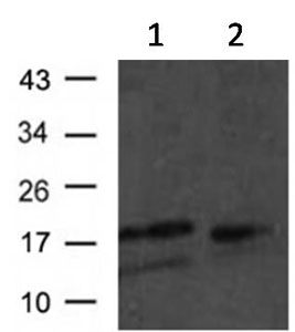
Anti-MAP1LC3A antibody [166AT1234] - ab168803 from Abcam

Prices $384.00
Sizes 400 µl
Host Mouse
Clonality Monoclonal
Clone 166AT1234
All lanes : Anti-MAP1LC3A antibody [166AT1234] (ab168803) at 1/1000 dilutionLane 1 : Hela cell lysates treated with DMSO Lane 2 : Hela cell lysates treated with rapamycin Lane 3 : Hela cell lysates treated with bafilomycin Lysates/proteins at 12.5 µg per lane.

Anti-MAP1LC3A antibody [EP1528Y] - ab52628 from Abcam

Prices $397.00
Sizes 100 µl
Host Rabbit
Clonality Monoclonal
Clone EP1528Y
Anti-MAP1LC3A antibody [EP1528Y] (ab52628) at 1/50000 dilution + Fetal brain lysate at 10 µgSecondarygoat anti-rabbit HRP labelled at 1/2000 dilution

Anti-MAP1LC3A antibody [EP1983Y] - Autophagosome Marker - ab52768 from Abcam

Prices $387.00
Sizes 100 µl
Host Rabbit
Clonality Monoclonal
Clone EP1983Y
Overlay histogram showing SHSY-5Y cells stained with ab52768 (red line). The cells were fixed with 4% paraformaldehyde (10 min) and then permeabilized with 0.1% PBS-Tween for 20 min. The cells were then incubated in 1x PBS / 10% normal goat serum / 0.3M glycine to block non-specific protein-protein interactions followed by the antibody (ab52768, 1/1000 dilution) for 30 min at 22°C. The secondary antibody used was Alexa Fluor® 488 goat anti-rabbit IgG (H&L) (ab150077) at 1/2000 dilution for 30 min at 22°C. Isotype control antibody (black line) was rabbit IgG (monoclonal) (0.1μg/1x106 cells) used under the same conditions. Unlabelled sample (blue line) was also used as a control. Acquisition of >5,000 events were collected using a 20mW Argon ion laser (488nm) and 525/30 bandpass filter. This antibody gave a positive signal in SHSY-5Y cells fixed with 80% methanol (5 min)/permeabilized with 0.1% PBS-Tween for 20 min used under the same conditions.

Anti-MAP1LC3A antibody [EP1983Y] - Autophagosome Marker (Alexa Fluor® 488) - ab185036 from Abcam

Prices $433.00
Sizes 100 µl
Host Rabbit
Clonality Monoclonal
Clone EP1983Y
Overlay histogram showing HeLa cells stained with ab185036 (red line). The cells were fixed with 4% formaldehyde (10 min) and then permeabilized with 0.1% PBS-Tween for 20 min. The cells were then incubated in 1x PBS / 10% normal goat serum / 0.3M glycine to block non-specific protein-protein interactions followed by the antibody (ab185036, 1/50 dilution) for 30 min at 22°C. Isotype control antibody (black line) was rabbit IgG (monoclonal) Alexa Fluor® 488 used at the same concentration and conditions as the primary antibody. Unlabelled sample (blue line) was also used as a control.Acquisition of >5,000 events were collected using a 20mW Argon ion laser (488nm) and 525/30 bandpass filter.This antibody gave a positive signal in HeLa fixed with 80% methanol (5 min)/permeabilized with 0.1% PBS-Tween for 20 min used under the same conditions.

Anti-MAP1LC3A antibody [EPR1754] - ab134178 from Abcam

Prices $379.00
Sizes 100 µl
Host Rabbit
Clonality Monoclonal
Clone EPR1754
Anti-MAP1LC3A antibody [EPR1754] (ab134178) at 1/500 dilution + HeLa cell lysate at 10 µgSecondaryHRP labelled goat anti-rabbit at 1/2000 dilution

Anti-MAP1LC3A antibody (HRP) - ab191810 from Abcam

Prices $359.00
Sizes 50 µg
Host Rabbit
Clonality Polyclonal
Search more