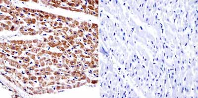
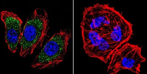
| Gene Symbol |
AP2A2
|
| Entrez Gene |
515216
|
| Alt Symbol |
-
|
| Species |
Bovine
|
| Gene Type |
protein-coding
|
| Description |
adaptor-related protein complex 2, alpha 2 subunit
|
| Other Description |
100 kDa coated vesicle protein C|AP-2 complex subunit alpha-2|adapter-related protein complex 2 alpha-2 subunit|adapter-related protein complex 2 subunit alpha-2|adaptor protein complex AP-2 subunit alpha-2|adaptor-related protein complex 2 subunit alpha-2|alpha-adaptin C|alpha2-adaptin|clathrin assembly protein complex 2 alpha-C large chain|plasma membrane adaptor HA2/AP2 adaptin alpha C subunit
|
| Swissprots |
Q0VCK5
|
| Accessions |
DAA13547 Q0VCK5 BC120121 AAI20122 NM_001075702 NP_001069170
|
| Function |
Component of the adaptor protein complex 2 (AP-2). Adaptor protein complexes function in protein transport via transport vesicles in different membrane traffic pathways. Adaptor protein complexes are vesicle coat components and appear to be involved in cargo selection and vesicle formation. AP-2 is involved in clathrin-dependent endocytosis in which cargo proteins are incorporated into vesicles surrounded by clathrin (clathrin- coated vesicles, CCVs) which are destined for fusion with the early endosome. The clathrin lattice serves as a mechanical scaffold but is itself unable to bind directly to membrane components. Clathrin-associated adaptor protein (AP) complexes which can bind directly to both the clathrin lattice and to the lipid and protein components of membranes are considered to be the major clathrin adaptors contributing the CCV formation. AP-2 also serves as a cargo receptor to selectively sort the membrane proteins involved in receptor-mediated endocytosis. AP-2 seems to p
|
| Subcellular Location |
Cell membrane {ECO:0000250}. Membrane, coated pit {ECO:0000250}; Peripheral membrane protein {ECO:0000250}; Cytoplasmic side {ECO:0000250}. Note=AP-2 appears to be excluded from internalizing CCVs and to disengage from sites of endocytosis seconds before internalization of the nascent CCV. {ECO:0000250}.
|
| Top Pathways |
Huntington's disease, Endocytosis, Endocrine and other factor-regulated calcium reabsorption, Synaptic vesicle cycle
|

