ATP5B

Gene Symbol ATP5B
Entrez Gene 327675
Alt Symbol -
Species Bovine
Gene Type protein-coding
Description ATP synthase, H+ transporting, mitochondrial F1 complex, beta polypeptide
Other Description ATP synthase subunit beta, mitochondrial|F1-ATPase beta-subunit|mitochondrial ATP synthase beta subunit
Swissprots Q9T2U5 Q1JQA9 P00829 Q9T2U4
Accessions DAA29742 P00829 BC116099 AAI16100 DQ403115 ABD77248 M20929 AAA30395 X05605 CAA29094 NM_175796 NP_786990
Function Mitochondrial membrane ATP synthase (F(1)F(0) ATP synthase or Complex V) produces ATP from ADP in the presence of a proton gradient across the membrane which is generated by electron transport complexes of the respiratory chain. F-type ATPases consist of two structural domains, F(1) - containing the extramembraneous catalytic core, and F(0) - containing the membrane proton channel, linked together by a central stalk and a peripheral stalk. During catalysis, ATP synthesis in the catalytic domain of F(1) is coupled via a rotary mechanism of the central stalk subunits to proton translocation. Subunits alpha and beta form the catalytic core in F(1). Rotation of the central stalk against the surrounding alpha(3)beta(3) subunits leads to hydrolysis of ATP in three separate catalytic sites on the beta subunits.
Subcellular Location Mitochondrion. Mitochondrion inner membrane. Note=Peripheral membrane protein.
Top Pathways Alzheimer's disease, Huntington's disease, Oxidative phosphorylation

Anti-ATPB antibody - ab128743 from Abcam

Prices $387.00
Sizes 100 µg
Host Rabbit
Clonality Polyclonal
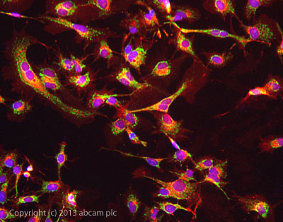
ab128743 stained HepG2 cells. The cells were 100% methanol fixed (5 min) and then incubated in 1%BSA / 10% normal goat serum / 0.3M glycine in 0.1% PBS-Tween for 1h to permeabilise the cells and block non-specific protein-protein interactions. The cells were then incubated with the antibody ab128743 at 5µg/ml overnight at +4°C. The secondary antibody (green) was DyLight® 488 goat anti- rabbit (ab96899) IgG (H+L) used at a 1/250 dilution for 1h. Alexa Fluor® 594 WGA was used to label plasma membranes (red) at a 1/200 dilution for 1h. DAPI was used to stain the cell nuclei (blue) at a concentration of 1.43µM.

Anti-ATPB antibody [3D5] - Mitochondrial Marker - ab14730 from Abcam

Prices $404.00
Sizes 100 µg
Host Mouse
Clonality Monoclonal
Clone 3D5
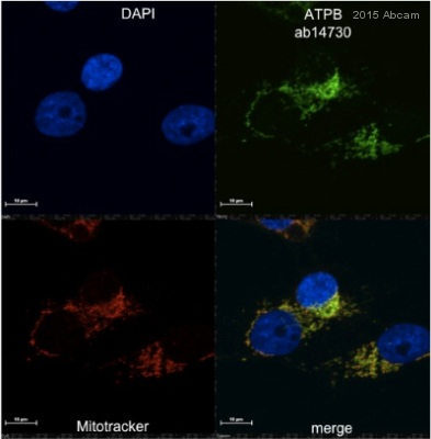
ab14730 staining ATPB in MDA MB 231 cells by ICC/IF (Immunocytochemistry/immunofluorescence). Cells were fixed with formaldehyde, permeabilized with 1% Triton X-100 and blocked with 10% BSA for 1 hour at 21°C. Samples were incubated with primary antibody (1/100 in PBS + 0.02% Tween 20) for 1 hour at 21°C. A DyLight® 550-conjugated goat anti-mouse IgG polyclonal (1/500) was used as the secondary antibody.See Abreview

Anti-ATPB antibody [3D5] - Mitochondrial Marker (Alexa Fluor® 488) - ab197904 from Abcam

Prices $425.00
Sizes 100 µl
Host Mouse
Clonality Monoclonal
Clone 3D5
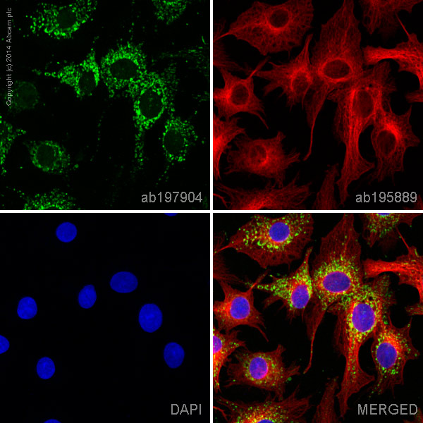
ab197904 staining ATPB in HepG2 cells. The cells were fixed with 4% formaldehyde (10 min), permeabilized with 0.1% Triton X-100 for 5 minutes and then blocked with 1% BSA/10% normal goat serum/0.3M glycine in 0.1% PBS-Tween for 1h. The cells were then incubated overnight at +4°C with ab197904 at a 1/100 dilution (shown in green) and ab195889, Mouse monoclonal to alpha Tubulin (Alexa Fluor® 594), at a 1/250 dilution (shown in red). Nuclear DNA was labelled with DAPI (shown in blue).Image was taken with a confocal microscope (Leica-Microsystems, TCS SP8).This product also gave a positive signal under the same testing conditions in HepG2 cells fixed with 100% methanol (5 min)

Anti-ATPB antibody [3D5] - Mitochondrial Marker (Alexa Fluor® 647) - ab197649 from Abcam

Prices $425.00
Sizes 100 µl
Host Mouse
Clonality Monoclonal
Clone 3D5
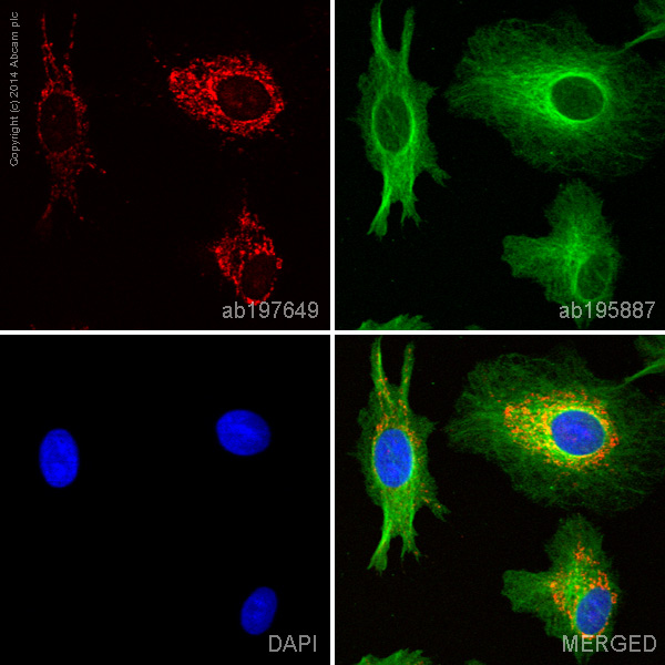
ab197649 staining ATPB in HepG2 cells. The cells were fixed with 100% methanol (5 min), permeabilized with 0.1% Triton X-100 for 5 minutes and then blocked with 1% BSA/10% normal goat serum/0.3M glycine in 0.1% PBS-Tween for 1h. The cells were then incubated overnight at +4°C with ab197649 at a 1/50 dilution (shown in red) and ab195887, Mouse monoclonal to alpha Tubulin (Alexa Fluor® 488), at a 1/250 dilution (shown in green). Nuclear DNA was labelled with DAPI (shown in blue).Image was taken with a confocal microscope (Leica-Microsystems, TCS SP8).

Anti-ATPB antibody [3D5] - Mitochondrial Marker (HRP) - ab197905 from Abcam

Prices $425.00
Sizes 100 µl
Host Mouse
Clonality Monoclonal
Clone 3D5
All lanes : Anti-ATPB antibody [3D5] - Mitochondrial Marker (HRP) (ab197905) at 1/5000 dilutionLane 1 : Human Heart Mitochondrial lysate (ab110337) at 5 µgLane 2 : HeLa (Human epithelial carcinoma cell line) Whole Cell Lysate (ab150035) at 10 µgLane 3 : HepG2 (Human hepatocellular liver carcinoma cell line) Whole Cell Lysate at 10 µgdeveloped using the ECL techniquePerformed under reducing conditions.

Anti-ATPB antibody [7E3F2] - ab110280 from Abcam

Prices $378.00
Sizes 100 µg
Host Mouse
Clonality Monoclonal
Clone 7E3F2
All lanes : Anti-ATPB antibody [7E3F2] (ab110280) at 4 µg/mlLane 1 : Isolated mitochondria from Human heart at 15 µgLane 2 : Isolated mitochondria from cow heart at 6 µgLane 3 : Isolated mitochondria from rat heart at 30 µgLane 4 : Isolated mitochondria from mouse heart at 30 µgLane 5 : Isolated mitochondria from HepG2 cells at 30 µg
Search more