PRKCB
| Gene Symbol | PRKCB |
|---|---|
| Entrez Gene | 5579 |
| Alt Symbol | PKC-beta, PKCB, PRKCB1, PRKCB2 |
| Species | Human |
| Gene Type | protein-coding |
| Description | protein kinase C, beta |
| Other Description | PKC-B|protein kinase C beta type|protein kinase C, beta 1 polypeptide |
| Swissprots | Q9UE50 D3DWF5 Q15138 C5IFJ8 O43744 Q9UEH8 P05127 Q9UE49 Q93060 Q9UJ33 Q9UJ30 P05771 |
| Accessions | AAA60096 AAA60097 AAB97933 AAB97934 AAD13852 ACS14045 BAA00912 CAA05725 CAA29395 CAA29396 CAA44393 CAZ00742 EAW55796 EAW55797 EAW55798 EAW55799 P05771 AA613106 AI423262 AJ002788 AK057555 AK123381 AL833252 BC036472 AAH36472 BC045175 BI549165 DB456157 EU832791 ACE87802 GQ129253 ACT64387 M13975 AAA60095 X06318 CAA29634 X07109 CAA30130 NM_002738 NP_002729 NM_212535 NP_997700 |
| Function | Calcium-activated, phospholipid- and diacylglycerol (DAG)-dependent serine/threonine-protein kinase involved in various cellular processes such as regulation of the B-cell receptor (BCR) signalosome, oxidative stress-induced apoptosis, androgen receptor-dependent transcription regulation, insulin signaling and endothelial cells proliferation. Plays a key role in B-cell activation by regulating BCR-induced NF-kappa-B activation. Mediates the activation of the canonical NF-kappa-B pathway (NFKB1) by direct phosphorylation of CARD11/CARMA1 at 'Ser-559', 'Ser-644' and 'Ser-652'. Phosphorylation induces CARD11/CARMA1 association with lipid rafts and recruitment of the BCL10-MALT1 complex as well as MAP3K7/TAK1, which then activates IKK complex, resulting in nuclear translocation and activation of NFKB1. Plays a direct role in the negative feedback regulation of the BCR signaling, by down-modulating BTK function via direct phosphorylation of BTK at 'Ser-180', which results in the alteration |
| Subcellular Location | Cytoplasm {ECO:0000250}. Nucleus {ECO:0000269|PubMed:20228790}. Membrane {ECO:0000250}; Peripheral membrane protein {ECO:0000250}. |
| Top Pathways | Wnt signaling pathway, Tight junction, VEGF signaling pathway, Focal adhesion, Gap junction |
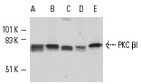
PKC βI (C-16) - sc-209 from Santa Cruz Biotechnology
|
||||||||||
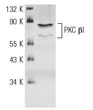
PKC βI (E-3) - sc-8049 from Santa Cruz Biotechnology
|
||||||||||
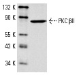
PKC βII (C-18) - sc-210 from Santa Cruz Biotechnology
|
||||||||||
PKC βII (F-7) - sc-13149 from Santa Cruz Biotechnology

|
||||||||||
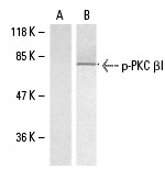
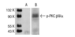
p-PKC βI (Thr 641) - sc-101776 from Santa Cruz Biotechnology
|
||||||||||
p-PKC βII/δ (Ser 660) - sc-11760 from Santa Cruz Biotechnology
|
||||||||||
Anti-PKC beta 1 + PKC beta 2 (phospho T500) antibody - ab5817 from Abcam
![Peptide Competition and Phosphatase Treatment: Lysates prepared from K562 cells stimulated with PMA were resolved by SDS-PAGE on a 10% polyacrylamide gel and transferred to PVDF. Membranes were either left untreated (1-4) or treated with lambda phosphatase (5), blocked with a 5% BSA-TBST buffer for one hour at room temperature, and incubated with ab5817 antibody for two hours at room temperature in a 3% BSA-TBST buffer, following prior incubation with: no peptide (1, 5), the non-phosphopeptide corresponding to the immunogen (2), a generic phosphothreonine-containing peptide (3), or, the phosphopeptide immunogen (4). After washing, membranes were incubated with goat F(ab’ 2 anti-rabbit IgG HRP conjugate and bands were detected using the Pierce SuperSignalTM method. The data show that only the peptide corresponding to PKC beta 1 & 2 [pT500] blocks the antibody signal. The data also show that phosphatase stripping eliminates the signal, verifying that the antibody is phospho-specific.](http://www.bioprodhub.com/system/product_images/ab_products/2/sub_4/12503_ab5817_1.jpg)
|
||||||||||
Anti-PKC beta 1 + PKC beta 2 (phospho T641) antibody - ab194749 from Abcam
|
||||||||||
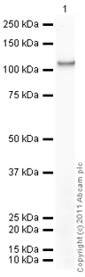
Anti-PKC beta 1 + PKC beta 2 antibody - ab5627 from Abcam
|
||||||||||
Anti-PKC beta 1 + PKC beta 2 antibody - ab71123 from Abcam
|
||||||||||
Anti-PKC beta 1 + PKC beta 2 antibody - N-terminal - ab189782 from Abcam
|
||||||||||
Anti-PKC beta 1 (phospho S661) antibody - ab79307 from Abcam
|
||||||||||
Anti-PKC beta 1 (phospho S661) antibody - ab192184 from Abcam
|
||||||||||
Anti-PKC beta 1 (phospho T642) antibody - ab75657 from Abcam
|
||||||||||
Anti-PKC beta 1 (phospho T642) antibody - ab5782 from Abcam
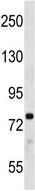
![Peptide Competition and Phosphatase Treatment: Lysates prepared from K562 cells stimulated with PMA were resolved by SDS-PAGE on a 10% polyacrylamide gel and transferred to PVDF. Membranes were either left untreated (1-4) or treated with lambda (ë) phosphatase (5), blocked with a 5% BSA-TBST buffer for one hour at room temperature, and incubated with ab5782 antibody for two hours at room temperature in a 3% BSA TBST buffer, following prior incubation with: no peptide (1, 5), the non phosphopeptide corresponding to the immunogen (2), a generic phosphothreonine-containing peptide (3), or, the phosphopeptide immunogen (4). After washing, membranes were incubated with goat F(ab’ 2 anti-rabbit IgG HRP conjugate and bands were detected using the Pierce SuperSignalTM method. The data show that only the peptide corresponding to PKC beta I [pT642] blocks the antibody signal. The data also show that phosphatase stripping eliminates the signal, verifying that the antibody is phospho-specific.](http://www.bioprodhub.com/system/product_images/ab_products/2/sub_4/12516_ab5782_1.jpg)
|
||||||||||
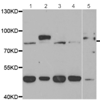
Anti-PKC beta 1 antibody - ab4132 from Abcam
|
||||||||||
Anti-PKC beta 1 antibody - ab137935 from Abcam
|
||||||||||
Anti-PKC beta 1 antibody [A10-F] - ab136917 from Abcam
|
||||||||||
Anti-PKC beta 1 antibody - C-terminal - ab175446 from Abcam
|
||||||||||
Anti-PKC beta 1 antibody - N-terminal - ab172919 from Abcam
|
||||||||||
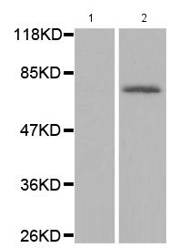
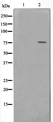
Anti-PKC beta 2 (phospho S660) antibody [EP1902Y] - ab75837 from Abcam
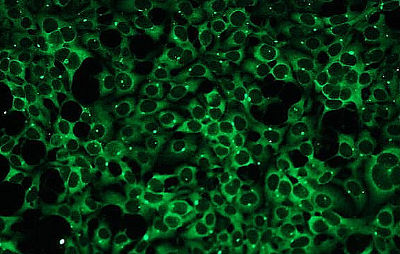
![All lanes : Anti-PKC beta 2 (phospho S660) antibody [EP1902Y] (ab75837) at 1/100000 dilutionLane 1 : Jurkat cell lysates, untreatedLane 2 : Jurkat cell lysates, treated with APLysates/proteins at 10 µg per lane.Secondarygoat anti-rabbit HRP at 1/2000 dilution](http://www.bioprodhub.com/system/product_images/ab_products/2/sub_4/12525_ab75837_1.JPG)
|
||||||||||
Anti-PKC beta 2 antibody - ab4138 from Abcam
|
||||||||||
Anti-PKC beta 2 antibody - ab4136 from Abcam
|
||||||||||
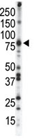
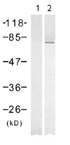
Anti-PKC beta 2 antibody [Y125] - ab32026 from Abcam
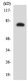
![Anti-PKC beta 2 antibody [Y125] (ab32026) at 1/500 dilution + K562 cell lysate](http://www.bioprodhub.com/system/product_images/ab_products/2/sub_4/12527_ab32026_1.JPG)
|
||||||||||
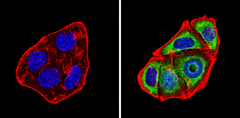
Anti-PKC beta 2 antibody [Y125] (Alexa Fluor® 488) - ab195251 from Abcam

|