SP1

Gene Symbol SP1
Entrez Gene 6667
Alt Symbol -
Species Human
Gene Type protein-coding
Description Sp1 transcription factor
Other Description specificity protein 1|transcription factor Sp1
Swissprots Q9NY21 Q9H3Q5 E4Z9M7 Q9NR51 P08047 Q86TN8 Q9NYE7 G5E9M8
Accessions AAF75063 AAF78780 BAB13476 CAP12210 CCQ43200 EAW96698 EAW96699 EAW96700 P08047 AB209678 BAD92915 AF252284 AAF67726 AF255682 AAF78781 AI282745 AI561005 AJ272134 CAB75345 AK311995 BAG34933 AL442093 AW028976 BC012008 BC043224 AAH43224 BC062539 AAH62539 BC078158 BF434304 BF436290 BF671559 BM463205 BQ072120 BQ431320 BQ774060 CD104746 FJ949572 ACR22508 FN908228 CBM42955 J03133 AAA61154 XM_011538696 XP_011536998 NM_001251825 NP_001238754 NM_003109 NP_003100 NM_138473 NP_612482
Function Transcription factor that can activate or repress transcription in response to physiological and pathological stimuli. Binds with high affinity to GC-rich motifs and regulates the expression of a large number of genes involved in a variety of processes such as cell growth, apoptosis, differentiation and immune responses. Highly regulated by post-translational modifications (phosphorylations, sumoylation, proteolytic cleavage, glycosylation and acetylation). Binds also the PDGFR- alpha G-box promoter. May have a role in modulating the cellular response to DNA damage. Implicated in chromatin remodeling. Plays a role in the recruitment of SMARCA4/BRG1 on the c-FOS promoter. Plays an essential role in the regulation of FE65 gene expression. In complex with ATF7IP, maintains telomerase activity in cancer cells by inducing TERT and TERC gene expression. Isoform 3 is a stronger activator of transcription than isoform 1. Positively regulates the transcription of the core clock component ARNTL/
Subcellular Location Nucleus. Cytoplasm. Note=Nuclear location is governed by glycosylated/phosphorylated states. Insulin promotes nuclear location, while glucagon favors cytoplasmic location.
Tissue Specificity Up-regulated in adenocarcinomas of the stomach (at protein level). Isoform 3 is ubiquitously expressed at low levels. {ECO:0000269|PubMed:21798247}.
Top Pathways TGF-beta signaling pathway, Huntington's disease, Estrogen signaling pathway, Choline metabolism in cancer, Transcriptional misregulation in cancer

SP1 Antibody - 5931 from Cell Signaling Technology

Prices $246.00
Sizes 100 µl (10 western blots)
Host Rabbit
Clonality Polyclonal
Western blot analysis of extracts from RL7, SR, and Ramos cells using SP1 Antibody.

SP1 (D4C3) Rabbit mAb - 9389 from Cell Signaling Technology

Prices $246.00
Sizes 100 µl (10 western blots)
Host Rabbit
Clonality Monoclonal
Clone D4C3
Western blot analysis of extracts from various cell lines using SP1 (D4C3) Rabbit mAb.

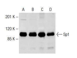
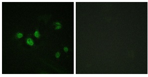
Sp1 (1C6) - sc-420 from Santa Cruz Biotechnology

Prices $279.00
Sizes 200 µg/ml
Host Mouse
Clonality Monoclonal
Sp1 (1C6): sc-420. Western blot analysis of Sp1 expression in K-562 (A,B) and Jurkat (C) nuclear extracts (A,C) and whole cell lysate (B).

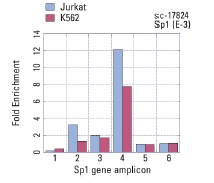
Sp1 (E-3) - sc-17824 from Santa Cruz Biotechnology

Prices $279.00
Sizes 200 µg/ml
Host Mouse
Clonality Monoclonal
Sp1 (E-3): sc-17824. ChIP analysis of Sp1 recruitment to genomic amplicons. Four (1-4) different human genomic Sp-1 and two control amplicons (5,6) were analyzed by quantitative PCR (primer sequences available in on-line supplemental data). Data generated in collaboration with Drs. N. Trinklein and R. Myers, Stanford University (ENCODE Project).

Sp1 (H-225) - sc-14027 from Santa Cruz Biotechnology

Prices $279.00
Sizes 200 µg/ml
Host Rabbit
Clonality Polyclonal
Sp1 (H-225): sc-14027. Immunoperoxidase staining of formalin fixed, paraffin-embedded human appendix tissue showing nuclear staining of glandular and lymphoid cells. Kindly provided by The Swedish Human Protein Atlas (HPA) program.

Sp1 (PEP 2) - sc-59 from Santa Cruz Biotechnology

Prices $279.00
Sizes 200 µg/ml
Host Rabbit
Clonality Polyclonal
Sp1 (PEP 2)-G: sc-59-G. Western blot analysis of Sp1 expression in K-562 (A) and Jurkat (C) nuclear extracts and K-562 (B) and Jurkat (D) whole cell lysates.

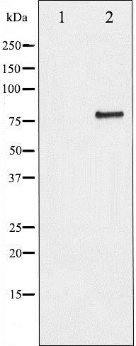
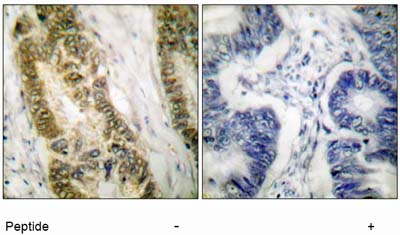
Anti-SP1 (phospho T453) antibody - ab37707 from Abcam

Prices $387.00
Sizes 100 µg
Host Rabbit
Clonality Polyclonal
All lanes : Anti-SP1 (phospho T453) antibody (ab37707) at 1 µg/mlLane 1 : HeLa (Human epithelial carcinoma cell line) Whole Cell LysateLane 2 : Jurkat (Human T cell lymphoblast-like cell line) Whole Cell LysateLane 3 : HeLa (Human epithelial carcinoma cell line) Whole Cell Lysate with Human SP1 (phospho T453) peptide (ab38350) at 1 µg/mlLane 4 : Jurkat (Human T cell lymphoblast-like cell line) Whole Cell Lysate with Human SP1 (phospho T453) peptide (ab38350) at 1 µg/mlLane 5 : HeLa (Human epithelial carcinoma cell line) Whole Cell Lysate with Human SP1 peptide (ab42654) at 1 µg/mlLane 6 : Jurkat (Human T cell lymphoblast-like cell line) Whole Cell Lysate with Human SP1 peptide (ab42654) at 1 µg/mlLysates/proteins at 10 µg per lane.SecondaryGoat polyclonal to Rabbit IgG - H&L - Pre-Adsorbed (HRP) at 1/3000 dilutiondeveloped using the ECL techniquePerformed under reducing conditions.

Anti-SP1 (phospho T453) antibody - ChIP Grade - ab59257 from Abcam

Prices $403.00
Sizes 100 µg
Host Rabbit
Clonality Polyclonal
Immunofluorescence analysis of HeLa cells, using ab59257 Antibody. The picture on the right is treated with the synthesized peptide.

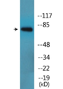
Anti-SP1 (phospho T739) antibody - ab195733 from Abcam

Prices $370.00
Sizes 100 µg
Host Rabbit
Clonality Polyclonal
All lanes : Anti-SP1 (phospho T739) antibody (ab195733) at 1/500 dilutionLane 1 : Serum treated COS7 whole cell lysate with antigen-specific peptideLane 2 : Serum treated COS7 whole cell lysateLysates/proteins at 12.5 µg per lane.

Anti-SP1 (phospho T739) antibody - ab138686 from Abcam

Host Rabbit
Clonality Polyclonal
Anti-SP1 (phospho T739) antibody (ab138686) at 1/500 dilution + COS7 cell extracts, treated with serum (20%, 15 mins) at 30 µg

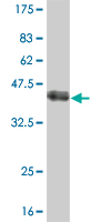
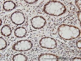
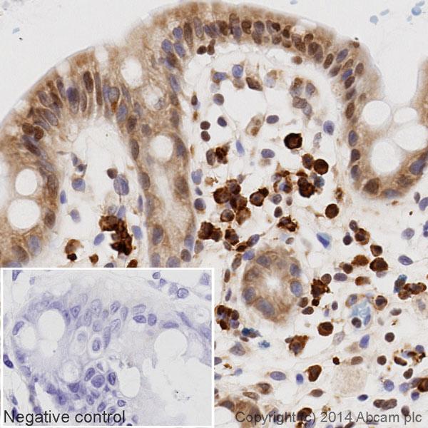
Anti-SP1 antibody - ab77441 from Abcam

Prices $398.00
Sizes 100 µg
Host Mouse
Clonality Monoclonal
Anti-SP1 antibody (ab77441) at 5 µg/ml + Recombinant fragment of human SP1 at 0.2 µgSecondaryGoat anti-Mouse IgG (H&L)-HRP at 1/5000 dilution

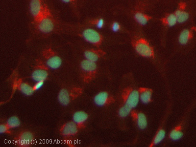
Anti-SP1 antibody - ab6082 from Abcam

Prices $385.00
Sizes 1 ml
Host Rabbit
Clonality Polyclonal
ICC/IF image of ab6082 stained HepG2 cells. The cells were 4% formaldehyde fixed (10 min) and then incubated in 1%BSA / 10% normal goat serum / 0.3M glycine in 0.1% PBS-Tween for 1h to permeabilise the cells and block non-specific protein-protein interactions. The cells were then incubated with the antibody (ab6082, 1µg/ml) overnight at +4°C. The secondary antibody (green) was Alexa Fluor® 488 goat anti-rabbit IgG (H+L) used at a 1/1000 dilution for 1h. Alexa Fluor® 594 WGA was used to label plasma membranes (red) at a 1/200 dilution for 1h. DAPI was used to stain the cell nuclei (blue) at a concentration of 1.43µM.

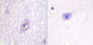
Anti-SP1 antibody - ab59267 from Abcam

Prices $379.00
Sizes 100 µg
Host Rabbit
Clonality Polyclonal
Immunohistochemical analysis of paraffin-embedded human brain tissue using ab59267 at a dilution of 1/50-1/100. Left hand image - without immunising peptide; right hand image - with immunising peptide.

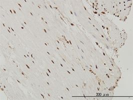
Anti-SP1 antibody - ab58202 from Abcam

Host Mouse
Clonality Monoclonal
SP1 antibody (ab58202) used in immunohistochemistry at 3ug/ml on formalin fixed and paraffin embedded human small Intestine.

Anti-SP1 antibody - ab52166 from Abcam

Host Rabbit
Clonality Polyclonal
Paraffin-embedded human colon carcinoma tissue stained for SP1 using ab52166 (1/100). The right-hand panel represents a negative control where ab52166 was pre-incubated with the immunizing (blocking) peptide.

Anti-SP1 antibody - ab28430 from Abcam

Host Rabbit
Clonality Polyclonal

Anti-SP1 antibody [1A5] - ab58199 from Abcam

Host Mouse
Clonality Monoclonal
Clone 1A5
SP1 antibody (ab58199) used in immunohistochemistry at 1ug/ml on formalin fixed and paraffin embedded human colon.

Anti-SP1 antibody [EPR6661] - ab133596 from Abcam

Prices $99.00, $387.00
Sizes 10 µl, 100 µl
Host Rabbit
Clonality Monoclonal
Clone EPR6661
All lanes : Anti-SP1 antibody [EPR6661] (ab133596) at 1/10000 dilution (purified)Lane 1 : A431 cell lysateLane 2 : HeLa cell lysateLane 3 : A549 cell lysateLysates/proteins at 20 µg per lane.SecondaryHRP goat anti-rabbit (H+L) at 1/1000 dilution

Anti-SP1 antibody [EPR6661] (Biotin) - ab197501 from Abcam

Prices $425.00
Sizes 100 µl
Host Rabbit
Clonality Monoclonal
Clone EPR6661
IHC image of SP1 staining in a section of formalin-fixed paraffin-embedded Hu_Colon_Norm*, performed on a Leica Bond system using the standard protocol B. The section was pre-treated using heat mediated antigen retrieval with sodium citrate buffer (pH6, epitope retrieval solution 1) for 20 mins. The section was then incubated with ab197501 at 1/100 dilution for 15 mins at room temperature and detected using an HRP conjugated ABC system. DAB was used as the chromogen. The section was then counterstained with haematoxylin and mounted with DPX. The inset negative control image is taken from an identical assay without primary antibody.For other IHC staining systems (automated and non-automated) customers should optimize variable parameters such as antigen retrieval conditions, primary antibody concentration and antibody incubation times.*Tissue obtained from the Human Research Tissue Bank, supported by the NIHR Cambridge Biomedical Research Centre

Anti-SP1 antibody [EPR6662(B)] - ab124804 from Abcam

Prices $99.00, $385.00
Sizes 10 µl, 100 µl
Host Rabbit
Clonality Monoclonal
Clone EPR6662(B)
All lanes : Anti-SP1 antibody [EPR6662(B)] (ab124804) at 1/1000 dilutionLane 1 : Ramos cell lysatesLane 2 : HeLa cell lysatesLane 3 : K562 cell lysatesLane 4 : Raji cell lysatesLysates/proteins at 10 µg per lane.

Anti-SP1 antibody [SP15H09] - ab51513 from Abcam

Prices $385.00
Sizes 100 µg
Host Mouse
Clonality Monoclonal
Clone SP15H09
Anti-SP1 antibody [SP15H09] (ab51513) at 1/100 dilution + HEK293 whole cell lysate at 25 µgSecondaryMouse IgG antibody at 1/2500 dilutiondeveloped using the ECL technique

Anti-SP1 Antibody (aa735-785) - LS-C290594 from LifeSpan Bioscience

Prices $375.00
Sizes 100 µl
Host Goat
Clonality Polyclonal
Detection of Human Sp1 by Immunohistochemistry. Sample: FFPE section of human ovarian carcinoma. Antibody: Affinity purified rabbit anti-Sp1 used at a dilution of 1:5000 (0.2 ug/ml). Detection: DAB.

Anti-SP1 Antibody (clone 1A5) IHC-plus™ - LS-B6148 from LifeSpan Bioscience

Prices $395.00
Sizes 50 µg
Host Mouse
Clonality Monoclonal
Clone 1A5
Anti-SP1 antibody IHC of human prostate. Immunohistochemistry of formalin-fixed, paraffin-embedded tissue after heat-induced antigen retrieval. Antibody LS-B6148 concentration 5 ug/ml.

Anti-SP1 Antibody (clone 1326CT463.109.176) - LS-C344150 from LifeSpan Bioscience

Prices $295.00
Sizes 400 µl
Host Mouse
Clonality Monoclonal
Clone 1326CT463.109.176
Fluorescent image of HeLa cells stained with SP1 Antibody. Antibody was diluted at 1:25 dilution. An Alexa Fluor 488-conjugated goat anti-mouse lgG at 1:400 dilution was used as the secondary antibody (green). Cytoplasmic actin was counterstained with Alexa Fluor 555 conjugated with Phalloidin (red).

Anti-SP1 Antibody (aa750-785) - LS-C39198 from LifeSpan Bioscience

Prices $680.00
Sizes 100 µg
Host Rabbit
Clonality Polyclonal
Search more