HSPD1

Gene Symbol HSPD1
Entrez Gene 424059
Alt Symbol -
Species Chicken
Gene Type protein-coding
Description heat shock 60kDa protein 1 (chaperonin)
Other Description 60 kDa chaperonin|60 kDa heat shock protein, mitochondrial|CPN60|HSP-60|chaperonin 60|heat shock protein 60|hsp60
Swissprots Q5ZL72 P84165
Accessions Q5ZL72 AJ719862 CAG31521 CR523558 NM_001012916 NP_001012934
Function Implicated in mitochondrial protein import and macromolecular assembly. May facilitate the correct folding of imported proteins. May also prevent misfolding and promote the refolding and proper assembly of unfolded polypeptides generated under stress conditions in the mitochondrial matrix (By similarity). {ECO:0000250|UniProtKB:P10809}.
Subcellular Location Mitochondrion matrix {ECO:0000250}.
Top Pathways RNA degradation

Anti-Hsp60 antibody - ab46798 from Abcam

Prices $403.00
Sizes 100 µl
Host Rabbit
Clonality Polyclonal
All lanes : Anti-Hsp60 antibody (ab46798) at 1/20000 dilution (purified)Lane 1 : Mouse brain tissue lysate at 20 µgLane 2 : Mouse heart tissue lysate at 20 µgLane 3 : Mouse spleen tissue lysate at 20 µgLane 4 : Rat heart tissue lysate at 10 µgSecondaryHRP conjugated goat anti-rabbit IgG (H+L) at 1/1000 dilution

Anti-Hsp60 antibody - ab137706 from Abcam

Prices $384.00
Sizes 100 µl
Host Rabbit
Clonality Polyclonal
All lanes : Anti-Hsp60 antibody (ab137706) at 1/10000 dilutionLane 1 : H1299 whole cell lysateLane 2 : HeLa S3 whole cell lysateLane 3 : MOLT4 whole cell lysateLysates/proteins at 30 µg per lane.

Anti-Hsp60 antibody - ab82520 from Abcam

Prices $376.00
Sizes 100 µg
Host Goat
Clonality Polyclonal
All lanes : Anti-Hsp60 antibody (ab82520) at 1/1000 dilutionLane 1 : Hsp60 Active Recombinant ProteinLane 2 : GroEL Active Recombinant ProteinLane 3 : Hsp65 ProteinLane 4 : HeLa Cell LysateLane 5 : 3T3 Cell LysateLane 6 : RK-13 Cell Lysate

Anti-Hsp60 antibody - ab109660 from Abcam

Prices $378.00
Sizes 100 µg
Host Rabbit
Clonality Polyclonal
All lanes : Anti-Hsp60 antibody (ab109660) at 1 µg/mlLane 1 : Recombinant Human Hsp60 at 0.1 µgLane 2 : SKBR3 lysate (Human) at 7.5 µgLane 3 : MDCK lysate (Dog) at 7.5 µgLane 4 : MEF lysate (Mouse) at 7.5 µg

Anti-Hsp60 antibody - ab31115 from Abcam

Prices $378.00
Sizes 500 µl
Host Rabbit
Clonality Polyclonal
All lanes : Anti-Hsp60 antibody (ab31115) at 1/8000 dilutionLane 1 : Cow normal mammary epithelial cells (nMEC) treated with serum free mediaLane 2 : Cow normal mammary epithelial cells (nMEC) treated with 1.0µM anisomycinLane 3 : Cow normal mammary epithelial cells (nMEC) treated with 100ng/ml IGF-1Lane 4 : Cow normal mammary epithelial cells (nMEC) treated with anisomycin and IGF-1Lane 5 : Cow normal mammary epithelial cells (nMEC) treated with serum free mediaLane 6 : Cow normal mammary epithelial cells (nMEC) treated with 1.0µM anisomycinLane 7 : Cow normal mammary epithelial cells (nMEC) treated with 100ng/ml IGF-1Lane 8 : Cow normal mammary epithelial cells (nMEC) treated with anisomycin and IGF-1Lysates/proteins at 40 µg per lane.SecondaryECL Anti-Rabbit IgG, Horseradish Peroxidase Linked at 1/20000 dilutiondeveloped using the ECL techniquePerformed under reducing conditions.

Anti-Hsp60 antibody - ab82513 from Abcam

Prices $376.00
Sizes 100 µg
Host Rabbit
Clonality Polyclonal
All lanes : Anti-Hsp60 antibody (ab82513) at 1/1000 dilutionLane 1 : HeLa (Heat Shocked) cell lysateLane 2 : 3T3 (Heat Shocked), cell lysateLane 3 : PC12 (Heat Shocked) cell lysate

Anti-Hsp60 antibody [2E1/53] - ab5479 from Abcam

Prices $388.00
Sizes 100 µg
Host Mouse
Clonality Monoclonal
Clone 2E1/53
Immunocytochemistry/Immunofluorescence analysis of Hsp60 (green) in Hela cells. Formalin-fixed cells were permeabilized with 0.1% Triton X-100 in TBS for 5-10 minutes at room temperature and blocked with 3% BSA-PBS for 30 minutes at room temperature. Cells were probed with ab5479 at a dilution of 1:200 and incubated overnight in a humidified chamber. Cells were washed with PBST and incubated with a DyLight-conjugated secondary antibody for 45 minutes at room temperature in the dark. F-actin (red) was stained with a fluorescent phalloidin and nuclei (blue) were stained with DAPI. Images were taken at a 60X magnification.

Anti-Hsp60 antibody [LK 1] - ab1819 from Abcam

Prices $384.00
Sizes 500 µl
Host Mouse
Clonality Monoclonal
Clone LK 1
ab1819 staining human breast carcinoma by IHC-P.

Anti-Hsp60 antibody [LK-1] - ab59457 from Abcam

Prices $403.00
Sizes 100 µg
Host Mouse
Clonality Monoclonal
Clone LK-1
ICC/IF image of ab59457 stained HepG2 cells. The cells were 100% methanol fixed (5 min) and then incubated in 1%BSA / 10% normal goat serum / 0.3M glycine in 0.1% PBS-Tween for 1h to permeabilise the cells and block non-specific protein-protein interactions. The cells were then incubated with the antibody (ab59457, 1µg/ml) overnight at +4°C. The secondary antibody (green) was Alexa Fluor® 488 goat anti-mouse IgG (H+L) used at a 1/1000 dilution for 1h. Alexa Fluor® 594 WGA was used to label plasma membranes (red) at a 1/200 dilution for 1h. DAPI was used to stain the cell nuclei (blue) at a concentration of 1.43µM.

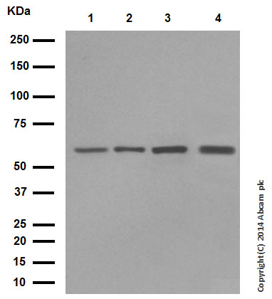
Anti-Hsp60 antibody [LK-1] - ab115879 from Abcam

Prices $385.00
Sizes 50 µg
Host Mouse
Clonality Monoclonal
Clone LK-1
All lanes : Anti-Hsp60 antibody [LK-1] (ab115879) at 1/1000 dilutionLane 1 : Hsp60 active recombinant proteinLane 2 : HeLa Cell lysate (heat shocked)Lane 3 : PC12 Cell lysate Lane 4 : NIH3T3 Cell lysate (heat shocked)

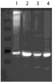
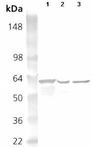
Anti-Hsp60 antibody [LK-2] - ab59458 from Abcam

Prices $385.00
Sizes 100 µg
Host Mouse
Clonality Monoclonal
Clone LK-2
Lanes 1 - 3 :  Anti-Hsp60 antibody [LK-2] (ab59458) at 1/4000 dilutionLane 4 : Lane 1 : Saccharomyces cerevisiae whole cell lysate at 6900000 cellsLane 2 : Saccharomyces cerevisiae whole cell lysate at 6900000 cellsLane 3 : Saccharomyces cerevisiae whole cell lysate at 6900000 cellsLane 4 : LadderSecondaryLanes 1 - 2 : HRP-conjugated goat anti-mouse IgG monoclonal at 1/5000 dilutionLane 3 : HRP-conjugated goat anti-mouse IgG monoclonal at 1/2000 dilutionPerformed under reducing conditions.

Anti-Hsp60 antibody [LK-2] (Phycoerythrin) - ab82518 from Abcam

Prices $376.00
Sizes 100 µg
Host Mouse
Clonality Monoclonal
Clone LK-2
ICC/IF image of ab82518 stained HeLa cells. The cells were 100% methanol fixed (5 min) and then incubated in 1%BSA / 10% normal goat serum / 0.3M glycine in 0.1% PBS-Tween for 1h to permeabilise the cells and block non-specific protein-protein interactions. The cells were then incubated with the antibody (ab82518, 10µg/ml) overnight at +4°C. The secondary antibody (green) was ab96879, DyLight® 488 goat anti-mouse IgG(H+L) used at a 1/250 dilution for 1h. Alexa Fluor® 594 WGA was used to label plasma membranes (red) at a 1/200 dilution for 1h. DAPI was used to stain the cell nuclei (blue) at a concentration of 1.43µM.

Anti-Hsp60 antibody [LK1] - ab3080 from Abcam

Host Mouse
Clonality Monoclonal
Clone LK1
Overlay histogram showing HeLa cells stained with ab3080 (red line). The cells were fixed with 80% methanol (5 min) and then permeabilized with 0.1% PBS-Tween for 20 min. The cells were then incubated in 1x PBS / 10% normal goat serum / 0.3M glycine to block non-specific protein-protein interactions followed by the antibody (ab3080, 0.1μg/1x106 cells) for 30 min at 22°C. The secondary antibody used was Alexa Fluor® 488 goat anti-mouse IgG (H+L) (ab150113) at 1/2000 dilution for 30 min at 22°C. Isotype control antibody (black line) was mouse IgG1 [ICIGG1] (ab91353, 1μg/1x106 cells) used under the same conditions. Unlabelled sample (blue line) was also used as a control. Acquisition of >5,000 events were collected using a 20mW Argon ion laser (488nm) and 525/30 bandpass filter.

Anti-Hsp60 antibody [LK1] - ab81333 from Abcam

Prices $379.00
Sizes 100 µl
Host Mouse
Clonality Monoclonal
Clone LK1
anti Hsp60 (form of ab81333 containing BSA and azide) at 2 µg/ml + human cells

Anti-Hsp60 antibody [LK1], prediluted - ab74184 from Abcam

Prices $378.00
Sizes 7 ml
Host Mouse
Clonality Monoclonal
Clone LK1
Formalin-fixed, paraffin-embedded human breast carcinoma stained with ab74184 using peroxidase-conjugate and AEC chromogen. Note intense cytoplasmic staining of tumor cells.
Search more