KIT

Gene Symbol KIT
Entrez Gene 3815
Alt Symbol C-Kit, CD117, PBT, SCFR
Species Human
Gene Type protein-coding
Description v-kit Hardy-Zuckerman 4 feline sarcoma viral oncogene homolog
Other Description mast/stem cell growth factor receptor Kit|p145 c-kit|piebald trait protein|proto-oncogene c-Kit|proto-oncogene tyrosine-protein kinase Kit|soluble KIT variant 1|tyrosine-protein kinase Kit|v-kit Hardy-Zuckerman 4 feline sarcoma viral oncogene-like protein
Swissprots Q99662 P10721 D5LXN2 B5A956 Q6IQ28 F5H8F8 D5M931 Q9UM99
Accessions AAB29301 AAB29529 AAC50968 AAC50969 ACI16424 ACI24886 CAA46771 CAA46772 CAA49159 CAJ15130 CAM35390 CAM91185 CAT16960 EAX05460 EAX05461 P10721 AJ438313 CAD27356 AK304031 BAG64945 BC071593 AAH71593 CN414753 DC376760 EU826594 ACF47630 GU983671 ADF36702 HM015525 ADF50068 HM015526 ADF50069 HM030712 ADF50070 HM030713 ADF50071 HM437240 ADN43071 X06182 CAA29548 XM_005265740 XP_005265797 XM_005265741 XP_005265798 XM_005265742 XP_005265799 NM_000222 NP_000213 NM_001093772 NP_001087241
Function Tyrosine-protein kinase that acts as cell-surface receptor for the cytokine KITLG/SCF and plays an essential role in the regulation of cell survival and proliferation, hematopoiesis, stem cell maintenance, gametogenesis, mast cell development, migration and function, and in melanogenesis. In response to KITLG/SCF binding, KIT can activate several signaling pathways. Phosphorylates PIK3R1, PLCG1, SH2B2/APS and CBL. Activates the AKT1 signaling pathway by phosphorylation of PIK3R1, the regulatory subunit of phosphatidylinositol 3-kinase. Activated KIT also transmits signals via GRB2 and activation of RAS, RAF1 and the MAP kinases MAPK1/ERK2 and/or MAPK3/ERK1. Promotes activation of STAT family members STAT1, STAT3, STAT5A and STAT5B. Activation of PLCG1 leads to the production of the cellular signaling molecules diacylglycerol and inositol 1,4,5-trisphosphate. KIT signaling is modulated by protein phosphatases, and by rapid internalization and degradation of the receptor. Activated KIT p
Subcellular Location Isoform 3: Cytoplasm. Note=Detected in the cytoplasm of spermatozoa, especially in the equatorial and subacrosomal region of the sperm head.
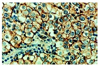
Tissue Specificity Isoform 1 and isoform 2 are detected in spermatogonia and Leydig cells. Isoform 3 is detected in round spermatids, elongating spermatids and spermatozoa (at protein level). Widely expressed. Detected in the hematopoietic system, the gastrointestinal system, in melanocytes and in germ cells. {ECO:0000269|PubMed:20601678, ECO:0000269|PubMed:2448137}.
Top Pathways Rap1 signaling pathway, Cytokine-cytokine receptor interaction, Central carbon metabolism in cancer, PI3K-Akt signaling pathway, Endocytosis

c-Kit (D13A2) XP ® Rabbit mAb (Biotinylated) - 5749 from Cell Signaling Technology

Prices $329.00
Sizes 100 µl (10 western blots)
Host Rabbit
Clonality Monoclonal
Clone D13A2
Western blot analysis of extracts from NCI-H526 cells using c-Kit (D13A2) XP ® Rabbit mAb (Biotinylated).

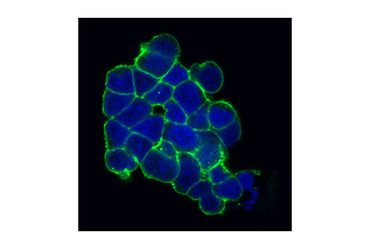
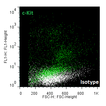
c-Kit (Ab81) Mouse mAb (Alexa Fluor ® 488 Conjugate) - 3310 from Cell Signaling Technology

Prices $299.00
Sizes 100 µl (50 tests)
Host Mouse
Clonality Monoclonal
Clone Ab81
Confocal immunofluorescent analysis of H526 cells using c-Kit (Ab81) Mouse mAb (Alexa Fluor ® 488 Conjugate) (green). Blue pseudocolor = DRAQ5 ® #4084 (fluorescent DNA dye).

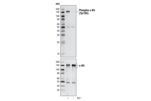
Phospho-c-Kit (Tyr703) (D12E12) Rabbit mAb - 3073 from Cell Signaling Technology

Prices $109.00, $287.00
Sizes 20 µl (2 western blots), 100 µl (10 western blots)
Host Rabbit
Clonality Monoclonal
Clone D12E12
Western blot analysis of extracts of H526 cells, untreated or treated with SCF, using Phospho-c-Kit (Tyr703) (D12E12) Rabbit mAb (upper) or c-Kit Antibody #3392 (lower).

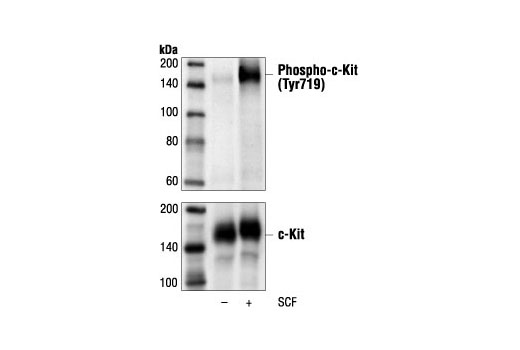
Phospho-c-Kit (Tyr719) Antibody - 3391 from Cell Signaling Technology

Prices $109.00, $287.00, $678.00
Sizes 20 µl (2 western blots), 100 µl (10 western blots), 300 µl (30 western blots)
Host Rabbit
Clonality Polyclonal
Western blot analysis of extracts from MO7e cells, untreated or stimulated with SCF #9907 (50 ng/ml for 2 minutes), using Phospho-c-Kit (Tyr719) Antibody (upper) or c-Kit Antibody #3392 (lower).

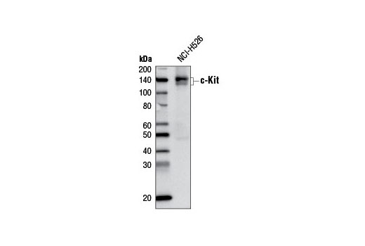
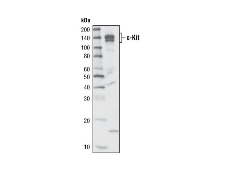
c-Kit (D13A2) XP ® Rabbit mAb - 3074 from Cell Signaling Technology

Prices $109.00, $277.00
Sizes 20 µl (2 western blots), 100 µl (10 western blots)
Host Rabbit
Clonality Monoclonal
Clone D13A2
Western blot analysis of extracts from NCI-H526 cells using c-Kit (D13A2) XP ® Rabbit mAb.

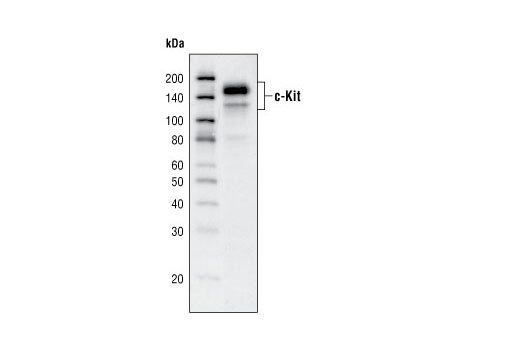
c-Kit (Ab81) Mouse mAb - 3308 from Cell Signaling Technology

Prices $246.00
Sizes 100 µl (10 western blots)
Host Mouse
Clonality Monoclonal
Clone Ab81
Western blot analysis of H526 cell lysate, using c-Kit (Ab81) Mouse mAb.

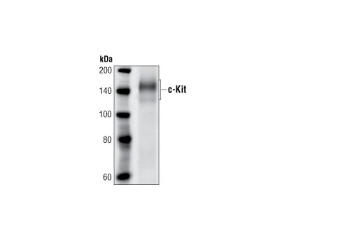
c-Kit Antibody - 3392 from Cell Signaling Technology

Prices $246.00
Sizes 100 µl (10 western blots)
Host Rabbit
Clonality Polyclonal
Western blot analysis of an extract from MO7e cells using c-Kit Antibody.

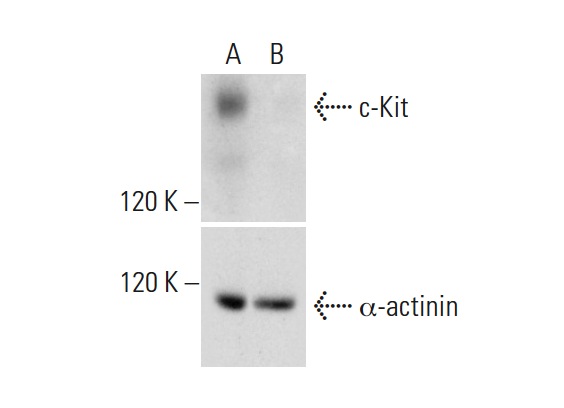
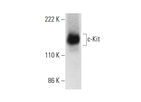
c-Kit (E-1) - sc-17806 from Santa Cruz Biotechnology

Prices $279.00
Sizes 200 µg/ml
Host Mouse
Clonality Monoclonal
c-Kit CRISPR/Cas9 KO Plasmid (h): sc-400106. Western blot analysis of human c-Kit expression in empty vector control (A) and c-Kit CRISPR/Cas9 KO Plasmid transfected (B) 293T whole cell lysates. Blot probed with c-Kit (E-1): sc-17806. α-actinin (H-2): sc-17829 used as specificity and loading control.

c-Kit (E-3) - sc-365504 from Santa Cruz Biotechnology

Prices $279.00
Sizes 200 µg/ml
Host Mouse
Clonality Monoclonal
c-Kit (E-3): sc-365504. Western blot analysis of c-Kit expression in HEL 92.1.7 whole cell lysate.

c-Kit (H-10) - sc-393910 from Santa Cruz Biotechnology

Prices $279.00
Sizes 200 µg/ml
Host Mouse
Clonality Monoclonal
c-Kit (H-10): sc-393910. Western blot analysis of c-Kit expression in CCRF-HSB-2 whole cell lysate.

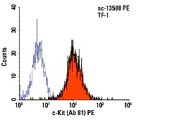
c-Kit (Ab 81) - sc-13508 from Santa Cruz Biotechnology

Prices $279.00
Sizes 200 µg/ml
Host Mouse
Clonality Monoclonal
c-Kit (Ab 81) PE: sc-13508 PE. FCM analysis of TF-1 cells. Black line histogram represents the isotype control, normal mouse IgG<sub>1</sub>: sc-2866.

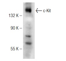
c-Kit (C-14) - sc-1493 from Santa Cruz Biotechnology

Prices $279.00
Sizes 200 µg/ml
Host Goat
Clonality Polyclonal
Western blot analysis of c-Kit expression in CCRF-HSB-2 whole cell lysates (A-C). Antibodies tested include c-Kit (C-19): sc-168 (A), c-Kit (C-14): sc-1493 (B) and c-Kit (M-14): sc-1494 (C).

c-Kit (H-300) - sc-5535 from Santa Cruz Biotechnology

Prices $279.00
Sizes 200 µg/ml
Host Rabbit
Clonality Polyclonal
c-Kit (H-300): sc-5535. Western blot analysis of c-Kit expression in CCRF-HSB-2 whole cell lysate.

c-Kit (C-19) - sc-168 from Santa Cruz Biotechnology

Prices $279.00
Sizes 200 µg/ml
Host Rabbit
Clonality Polyclonal
c-Kit (C-19): sc-168. Immunoperoxidase staining of Bouins-fixed, paraffin-embedded invasive testicular germ cell tumor tissue showing distinct membrane staining. Kindly provided by E. Rajpert-De Meyts.

c-Kit (M-14) - sc-1494 from Santa Cruz Biotechnology

Prices $279.00
Sizes 200 µg/ml
Host Goat
Clonality Polyclonal
Western blot analysis of c-Kit expression in CCRF-HSB-2 whole cell lysates (A-C). Antibodies tested include c-Kit (C-19): sc-168 (A), c-Kit (C-14): sc-1493 (B) and c-Kit (M-14): sc-1494 (C).

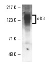
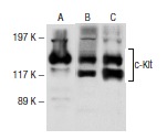
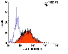
c-Kit (104D2) - sc-19983 from Santa Cruz Biotechnology

Prices $279.00
Sizes 200 µg/ml
Host Mouse
Clonality Monoclonal
c-Kit (104D2) PE: sc-19983 PE. FCM analysis of TF-1 cells. Black line histogram represents the isotype control, normal mouse IgG<sub>1</sub>: sc-2866.

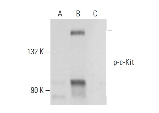
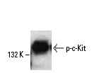
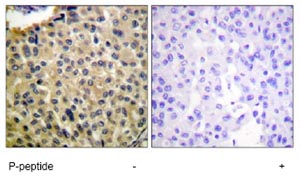
p-c-Kit (Tyr 568/570) - sc-18076 from Santa Cruz Biotechnology

Prices $279.00
Sizes 200 µg/ml
Host Goat
Clonality Polyclonal
p-c-Kit (Tyr 568/570)-R: sc-18076-R. Western blot analysis of c-Kit phosphorylation in untreated (A), EGF treated (B) and EGF and lambda protein phosphatase treated (C) A-431 whole cell lysates.

p-c-Kit (Tyr 721) - sc-101659 from Santa Cruz Biotechnology

Prices $279.00
Sizes 100 µg/ml
Host Rabbit
Clonality Polyclonal
p-c-Kit (Tyr 721): sc-101659. Western blot analysis of phosphorylated c-Kit expression in SCF-treated 293 transfected whole cell lysate.

Anti-c-Kit (phospho Y568 + Y570) antibody - ab5616 from Abcam

Prices $370.00
Sizes 50 µl
Host Rabbit
Clonality Polyclonal
ab5616 staining c-Kit (phospho Y568 + Y570) in human NCI-60 cells by Flow Cytometry. Cells were fixed with paraformaldehyde and permeabilized. The sample was incubated with the primary antibody (1/200) for 1 hour at 20°C. An Alexa Fluor® 488-conjugated goat anti-rabbit IgG polyclonal (1/1000) was used as the secondary antibody.Gating Strategy: isotype population (shown in white).See Abreview

Anti-c-Kit (phospho Y703) antibody - ab62154 from Abcam

Prices $370.00
Sizes 100 µg
Host Rabbit
Clonality Polyclonal
Immunohistochemistry analysis of paraffin-embedded human breast carcinoma tissue using c-Kit (phospho Y703) antibody (ab62154) at 1/50 - 1/100 dilution, in the presence (right panel) and absence (left panel) of immunising phosphopeptide.

Anti-c-Kit (phospho Y703) antibody - ab195737 from Abcam

Prices $370.00
Sizes 100 µg
Host Rabbit
Clonality Polyclonal
All lanes : Anti-c-Kit (phospho Y703) antibody (ab195737) at 1/500 dilutionLane 1 : EGF treated HepG2 whole cell lysate with antigen-specific peptideLane 2 : EGF treated HepG2 whole cell lysateLysates/proteins at 12.5 µg per lane.

Anti-c-Kit (phospho Y721) antibody - ab195741 from Abcam

Prices $370.00
Sizes 100 µg
Host Rabbit
Clonality Polyclonal
All lanes : Anti-c-Kit (phospho Y721) antibody (ab195741) at 1/500 dilutionLane 1 : HeLa whole cell lysate with antigen-specific peptideLane 2 : HeLa whole cell lysateLysates/proteins at 12.5 µg per lane.

Anti-c-Kit (phospho Y721) antibody - ab47766 from Abcam

Prices $370.00
Sizes 100 µg
Host Rabbit
Clonality Polyclonal
All lanes : Anti-c-Kit (phospho Y721) antibody (ab47766) at 1/500 dilutionLane 1 : MO7e cell extract, treated with SCF (50 ng/ml for 2 min) Lane 2 : MO7e cell extract, untreated

Anti-c-Kit (phospho Y936) antibody - ab138433 from Abcam

Prices $370.00
Sizes 100 µg
Host Rabbit
Clonality Polyclonal
Anti-c-Kit (phospho Y936) antibody (ab138433) at 1/500 dilution + extracts from HepG2 cells treated with 200 ng/ml EGF for 30 minutes

Anti-c-Kit antibody - ab5505 from Abcam

Prices $384.00
Sizes 400 µl
Host Rabbit
Clonality Polyclonal
ICC/IF image of ab5505 stained SKNSH cells. The cells were 4% formaldehyde fixed (10 min) and then incubated in 1%BSA / 10% normal goat serum/ 0.3M glycine in 0.1% PBS-Tween for 1h to permeabilise the cells and block non-specific protein-protein interactions. The cells were then incubated with the antibody ab5505 at 5 µg/mL overnight at +4°C. The secondary antibody (green) was DyLight® 488 Goat anti-Rabbit IgG (H+L) (ab96899) used at a 1/250 dilution for 1h. Alexa Fluor® 594 WGA was used to label plasma membranes (red) at a 1/200 dilution for 1h. DAPI was used to stain the cell nuclei (blue) at a concentration of 1.43µM.
Search more