CTSD

Gene Symbol CTSD
Entrez Gene 1509
Alt Symbol CLN10, CPSD, HEL-S-130P
Species Human
Gene Type protein-coding
Description cathepsin D
Other Description ceroid-lipofuscinosis, neuronal 10|epididymis secretory sperm binding protein Li 130P|lysosomal aspartyl peptidase|lysosomal aspartyl protease
Swissprots Q6IB57 P07339
Accessions AAA16314 AAA51922 AAD13868 AAD14156 EAX02461 EAX02462 EAX02463 P07339 AK130178 BC001574 BC016320 AAH16320 BM762986 BM976200 BT006910 AAP35556 BT020155 AAV38957 CR456947 CAG33228 DQ893547 ABM84473 DQ893878 ABM84804 EU794598 ACJ13652 M11233 AAB59529 X05344 CAA28955 NM_001909 NP_001900
Function Acid protease active in intracellular protein breakdown. Involved in the pathogenesis of several diseases such as breast cancer and possibly Alzheimer disease.
Subcellular Location Lysosome. Melanosome. Secreted, extracellular space. Note=Identified by mass spectrometry in melanosome fractions from stage I to stage IV. In aortic samples, detected as an extracellular protein loosely bound to the matrix (PubMed:20551380). {ECO:0000269|PubMed:20551380}.
Tissue Specificity Expressed in the aorta extrcellular space (at protein level). {ECO:0000269|PubMed:20551380}.
Top Pathways Lysosome, Tuberculosis, Sphingolipid signaling pathway

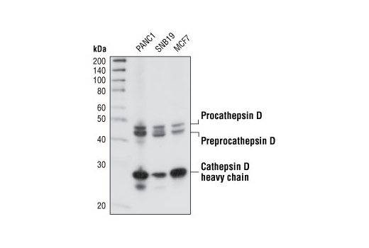
Cathepsin D Antibody - 2284 from Cell Signaling Technology

Prices $246.00
Sizes 100 µl (10 western blots)
Host Rabbit
Clonality Polyclonal
Western blot analysis of extracts from PANC1, SNB19 and MCF-7 cell lines using Cathepsin D Antibody.

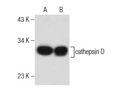
cathepsin D (C-5) - sc-377124 from Santa Cruz Biotechnology

Prices $279.00
Sizes 200 µg/ml
Host Mouse
Clonality Monoclonal
cathepsin D (C-5): sc-377124. Western blot analysis of cathepsin D expression in MCF7 (A) and ZR-75-1 (B) whole cell lysates.

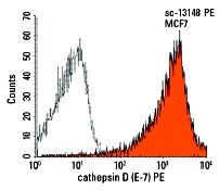
cathepsin D (E-7) - sc-13148 from Santa Cruz Biotechnology

Prices $279.00
Sizes 200 µg/ml
Host Mouse
Clonality Monoclonal
cathepsin D (E-7) PE: sc-13148 PE. Intracellular FCM analysis of fixed and permeabilized MCF7 cells. Black line histogram represents the isotype control, normal mouse IgG<sub>1</sub>: sc-2866.

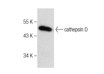
cathepsin D (D-7) - sc-377299 from Santa Cruz Biotechnology

Prices $279.00
Sizes 200 µg/ml
Host Mouse
Clonality Monoclonal
cathepsin D (D-7): sc-377299. Western blot analysis of cathepsin D expression in ZR-75-1 whole cell lysate.

cathepsin D (C-20) - sc-6486 from Santa Cruz Biotechnology

Prices $279.00
Sizes 200 µg/ml
Host Goat
Clonality Polyclonal
cathepsin D (C-20): sc-6486. Immunoperoxidase staining of formalin-fixed, paraffin-embedded human breast tumor showing cytoplasmic staining.

cathepsin D (F-12) - sc-374381 from Santa Cruz Biotechnology

Prices $279.00
Sizes 200 µg/ml
Host Mouse
Clonality Monoclonal
cathepsin D (F-12): sc-374381. Western blot analysis of cathepsin D expression in MCF7 (A) and ZR-75-1 (B) whole cell lysates.

cathepsin D (G-19) - sc-6494 from Santa Cruz Biotechnology

Prices $279.00
Sizes 200 µg/ml
Host Goat
Clonality Polyclonal
cathepsin D (G-19): sc-6494. Immunofluorescence staining of methanol-fixed ZR-75-1 cells showing punctated cytoplasmic/lysosomal staining.

cathepsin D (H-75) - sc-10725 from Santa Cruz Biotechnology

Prices $279.00
Sizes 200 µg/ml
Host Rabbit
Clonality Polyclonal
cathepsin D (H-75): sc-10725. Immunoperoxidase staining of formalin-fixed, paraffin-embedded human breast tumor showing cytoplasmic staining.

cathepsin D (N-19) - sc-6488 from Santa Cruz Biotechnology

Prices $279.00
Sizes 200 µg/ml
Host Goat
Clonality Polyclonal
cathepsin D (N-19): sc-6488. Immunofluorescence staining of methanol-fixed ZR-75-1 cells showing cytoplasmic staining.

cathepsin D (R-20) - sc-6487 from Santa Cruz Biotechnology

Prices $279.00
Sizes 200 µg/ml
Host Goat
Clonality Polyclonal
cathepsin D (R-20): sc-6487. Western blot analysis of cathepsin D precursors (upper, middle) and processed form (lower) expression in MCF7 (A) and ZR-75-1 (B) whole cell lysates and rat kidney extract (C).

cathepsin D (4G2) - sc-53927 from Santa Cruz Biotechnology

Prices $279.00
Sizes 100 µg/ml
Host Mouse
Clonality Monoclonal
cathepsin D (4G2): sc-53927. Western blot analysis of cathepsin D expression in Hep G2 (A), Hep 3B (B), MDA-MB-231 (C) and MCF7 (D) whole cell lysates.

cathepsin D (49) - sc-136282 from Santa Cruz Biotechnology

Prices $279.00
Sizes 50 µg/0.5 ml
Host Mouse
Clonality Monoclonal
cathepsin D (49): sc-136282. Western blot analysis of cathepsin D expression in Hep G2 whole cell lysate.

Anti-Cathepsin D antibody - ab826 from Abcam

Prices $385.00
Sizes 250 µl
Host Rabbit
Clonality Polyclonal
ICC/IF image of ab826 stained A431 cells. The cells were 100% methanol fixed (5 min) and then incubated in 1%BSA / 10% normal goat serum / 0.3M glycine in 0.1% PBS-Tween for 1h to permeabilise the cells and block non-specific protein-protein interactions. The cells were then incubated with the antibody (ab826, 10µg/ml) overnight at +4°C. The secondary antibody (green) was ab96899, DyLight® 488 goat anti-rabbit IgG (H+L) used at a 1/250 dilution for 1h. Alexa Fluor® 594 WGA was used to label plasma membranes (red) at a 1/200 dilution for 1h. DAPI was used to stain the cell nuclei (blue) at a concentration of 1.43µM.

Anti-Cathepsin D antibody - ab19555 from Abcam

Prices $379.00
Sizes 100 µl
Host Rabbit
Clonality Polyclonal
IHC image of Cathepsin D staining in Human Lung formalin fixed paraffin embedded tissue section, performed on a Leica Bond™ system using the standard protocol F. The section was pre-treated using heat mediated antigen retrieval with sodium citrate buffer (pH6, epitope retrieval solution 1) for 20 mins. The section was then incubated with ab19555, 1/2000 dilution, for 15 mins at room temperature and detected using an HRP conjugated compact polymer system. DAB was used as the chromogen. The section was then counterstained with haematoxylin and mounted with DPX. For other IHC staining systems (automated and non-automated) customers should optimize variable parameters such as antigen retrieval conditions, primary antibody concentration and antibody incubation times.

Anti-Cathepsin D antibody - ab72915 from Abcam

Prices $384.00
Sizes 100 µg
Host Rabbit
Clonality Polyclonal
IHC image of Cathepsin D staining in human adrenal gland formalin fixed paraffin embedded tissue section, performed on a Leica Bond system using the standard protocol F. The section was pre-treated using heat mediated antigen retrieval with sodium citrate buffer (pH6, epitope retrieval solution 1) for 20 mins. The section was then incubated with ab72915, 0.1µg/ml, for 15 mins at room temperature and detected using an HRP conjugated compact polymer system. DAB was used as the chromogen. The section was then counterstained with haematoxylin and mounted with DPX.For other IHC staining systems (automated and non-automated) customers should optimize variable parameters such as antigen retrieval conditions, primary antibody concentration and antibody incubation times.

Anti-Cathepsin D antibody - ab97499 from Abcam

Prices $376.00
Sizes 100 µl
Host Rabbit
Clonality Polyclonal
IHC image of Cathepsin D staining in Human Lung formalin fixed paraffin embedded tissue section, performed on a Leica Bond™ system using the standard protocol F. The section was pre-treated using heat mediated antigen retrieval with sodium citrate buffer (pH6, epitope retrieval solution 1) for 20 mins. The section was then incubated with ab97499, 0.5 µg/ml, for 15 mins at room temperature and detected using an HRP conjugated compact polymer system. DAB was used as the chromogen. The section was then counterstained with haematoxylin and mounted with DPX. For other IHC staining systems (automated and non-automated) customers should optimize variable parameters such as antigen retrieval conditions, primary antibody concentration and antibody incubation times.

Anti-Cathepsin D antibody - ab49787 from Abcam

Prices $378.00
Sizes 100 µg
Host Rabbit
Clonality Polyclonal
All lanes : Anti-Cathepsin D antibody (ab49787) at 1/1000 dilutionLane 1 : Recombinant Human Cathepsin D at 0.04 µgLane 2 : Recombinant Human Cathepsin D at 0.02 µgLane 3 : Recombinant Human Cathepsin D at 0.01 µgLane 4 : Recombinant Human Cathepsin D at 0.004 µgLane 5 : Recombinant Human Cathepsin D at 0.002 µg

Anti-Cathepsin D antibody - ab49788 from Abcam

Prices $376.00
Sizes 100 µg
Host Rabbit
Clonality Polyclonal
All lanes : Anti-Cathepsin D antibody (ab49788) at 1/1000 dilutionLane 1 : Recombinant Human Cathepsin D at 0.04 µgLane 2 : Recombinant Human Cathepsin D at 0.02 µgLane 3 : Recombinant Human Cathepsin D at 0.01 µgLane 4 : Recombinant Human Cathepsin D at 0.004 µgLane 5 : Recombinant Human Cathepsin D at 0.002 µg

Anti-Cathepsin D antibody - ab49789 from Abcam

Prices $376.00
Sizes 100 µg
Host Rabbit
Clonality Polyclonal
All lanes : Anti-Cathepsin D antibody (ab49789) at 1/1000 dilutionLane 1 : r-Human Cathepsin D at 0.04 µgLane 2 : r-Human Cathepsin D at 0.02 µgLane 3 : r-Human Cathepsin D at 0.01 µgLane 4 : r-Human Cathepsin D at 0.004 µgLane 5 : r-Human Cathepsin D at 0.002 µg

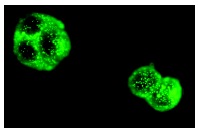
Anti-Cathepsin D antibody [4G2] - ab40697 from Abcam

Prices $398.00
Sizes 100 µl
Host Mouse
Clonality Monoclonal
Clone 4G2
Overlay histogram showing MCF7 cells stained with ab40697 (red line). The cells were fixed with 80% methanol (5 min) and then permeabilized with 0.1% PBS-Tween for 20 min. The cells were then incubated in 1x PBS / 10% normal goat serum / 0.3M glycine to block non-specific protein-protein interactions followed by the antibody (ab40697, 1μg/1x106 cells) for 30 min at 22°C. The secondary antibody used was Alexa Fluor® 488 goat anti-mouse IgG (H+L) (ab150113) at 1/2000 dilution for 30 min at 22°C. Isotype control antibody (black line) was mouse IgG2b [PLPV219] (ab91366, 1μg/1x106 cells) used under the same conditions. Unlabelled sample (blue line) was also used as a control. Acquisition of >5,000 events were collected using a 20mW Argon ion laser (488nm) and 525/30 bandpass filter.

Anti-Cathepsin D antibody [CTD-19] - ab6313 from Abcam

Prices $397.00
Sizes 100 µl
Host Mouse
Clonality Monoclonal
Clone CTD-19
ICC/IF image of ab6313 stained human HepG2 cells. The cells were 4% PFA fixed (10 min) and then incubated in 1%BSA / 10% normal goat serum / 0.3M glycine in 0.1% PBS-Tween for 1h to permeabilise the cells and block non-specific protein-protein interactions. The cells were then incubated with the antibody (ab6313, 5 µg/ml) overnight at +4°C. The secondary antibody (green) was Alexa Fluor® 488 goat anti-mouse IgG (H+L) used at a 1/1000 dilution for 1h. Alexa Fluor® 594 WGA was used to label plasma membranes (red) at a 1/200 dilution for 1h. DAPI was used to stain the cell nuclei (blue). This antibody also gave a positive IF result in HeLa, Hek293 and MCF7 cells.

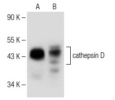
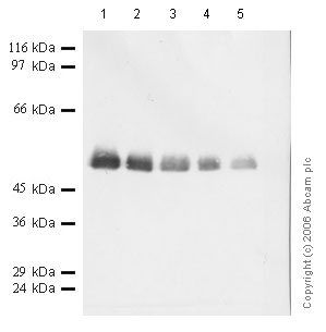
Anti-Cathepsin D antibody [EPR3054] - ab134169 from Abcam
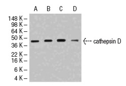
Prices $385.00
Sizes 100 µl
Host Rabbit
Clonality Monoclonal
Clone EPR3054
All lanes : Anti-Cathepsin D antibody [EPR3054] (ab134169) at 1/2000 dilution (purified)Lane 1 : MCF-7 cell lysateLane 2 : A431 cell lysateLane 3 : SKBR-3 cell lysateLysates/proteins at 20 µg per lane.SecondaryHRP goat anti-rabbit (H+L) at 1/1000 dilution

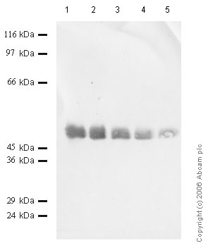
Anti-Cathepsin D antibody [EPR3056Y] - ab75811 from Abcam
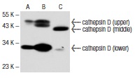
Prices $379.00
Sizes 100 µl
Host Rabbit
Clonality Monoclonal
Clone EPR3056Y
All lanes : Anti-Cathepsin D antibody [EPR3056Y] (ab75811) at 1/1000 dilutionLane 1 : MCF-7 cell lysateLane 2 : SKBR3 cell lysateLysates/proteins at 10 µg per lane.SecondaryHRP labelled goat anti-rabbit at 1/2000 dilution

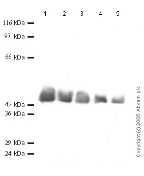
Anti-Cathepsin D antibody [EPR3057Y] - ab75852 from Abcam
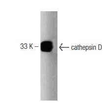
Prices $397.00
Sizes 100 µl
Host Rabbit
Clonality Monoclonal
Clone EPR3057Y
All lanes : Anti-Cathepsin D antibody [EPR3057Y] (ab75852) at 1/10000 dilutionLane 1 : MCF-7 cell lysateLane 2 : A431 cell lysateLane 3 : SKBR3 cell lysateLane 4 : HepG2 cell lysateLysates/proteins at 10 µg per lane.SecondaryHRP-labeled goat anti-rabbit at 1/2000 dilution

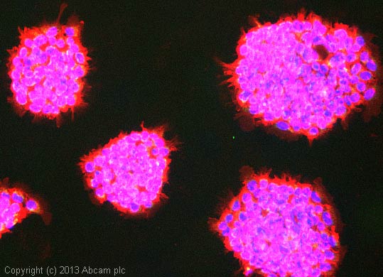
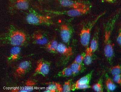
Anti-Cathepsin D antibody [EPR3057Y] (Alexa Fluor® 647) - ab198326 from Abcam
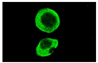
Prices $425.00
Sizes 100 µl
Host Rabbit
Clonality Monoclonal
Clone EPR3057Y
ab198326 staining Cathepsin D in MCF-7 cells. The cells were fixed with 100% methanol (5 min), permeabilised in 0.1% Triton X-100 for 5 minutes and then blocked in 1% BSA/10% normal goat serum/0.3M glycine in 0.1% PBS-Tween for 1h. The cells were then incubated with ab198326 at 1/100 dilution (shown in red) and ab195887, Mouse monoclonal [DM1A] to alpha Tubulin (Alexa Fluor® 488, shown in green) at 1/167 dilution overnight at +4°C. Nuclear DNA was labelled in blue with DAPI.Image was taken with a confocal microscope (Leica-Microsystems, TCS SP8).
Search more