CSPG4
| Gene Symbol | CSPG4 |
|---|---|
| Entrez Gene | 1464 |
| Alt Symbol | HMW-MAA, MCSP, MCSPG, MEL-CSPG, MSK16, NG2 |
| Species | Human |
| Gene Type | protein-coding |
| Description | chondroitin sulfate proteoglycan 4 |
| Other Description | chondroitin sulfate proteoglycan 4 (melanoma-associated)|chondroitin sulfate proteoglycan NG2|melanoma chondroitin sulfate proteoglycan|melanoma-associated chondroitin sulfate proteoglycan |
| Swissprots | Q6UVK1 Q92675 D3DW77 |
| Accessions | EAW99239 EAW99240 Q6UVK1 AY359468 AAQ62842 BC033194 BC128110 AAI28111 BC172270 AAI72270 BC172576 AAI72576 BP363063 BQ679485 CA428796 CA438367 CB044012 X96753 CAA65529 NM_001897 NP_001888 |
| Function | Proteoglycan playing a role in cell proliferation and migration which stimulates endothelial cells motility during microvascular morphogenesis. May also inhibit neurite outgrowth and growth cone collapse during axon regeneration. Cell surface receptor for collagen alpha 2(VI) which may confer cells ability to migrate on that substrate. Binds through its extracellular N- terminus growth factors, extracellular matrix proteases modulating their activity. May regulate MPP16-dependent degradation and invasion of type I collagen participating in melanoma cells invasion properties. May modulate the plasminogen system by enhancing plasminogen activation and inhibiting angiostatin. Functions also as a signal transducing protein by binding through its cytoplasmic C-terminus scaffolding and signaling proteins. May promote retraction fiber formation and cell polarization through Rho GTPase activation. May stimulate alpha-4, beta-1 integrin- mediated adhesion and spreading by recruiting and activat |
| Subcellular Location | Apical cell membrane {ECO:0000250}; Single- pass type I membrane protein {ECO:0000250}; Extracellular side {ECO:0000250}. Cell projection, lamellipodium membrane {ECO:0000250}; Single-pass type I membrane protein {ECO:0000250}; Extracellular side {ECO:0000250}. Note=Localized at the apical plasma membrane it relocalizes to the lamellipodia of astrocytoma upon phosphorylation by PRKCA. Localizes to the retraction fibers. Localizes to the plasma membrane of oligodendrocytes (By similarity). {ECO:0000250}. |
| Tissue Specificity | Detected only in malignant melanoma cells. {ECO:0000269|PubMed:8790396}. |
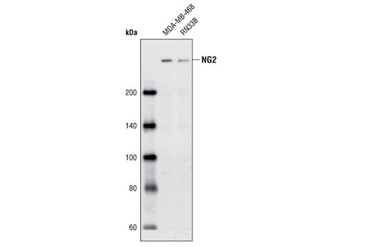
NG2 Antibody - 4235 from Cell Signaling Technology
|
||||||||||
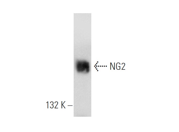
NG2 (D-20) - sc-30922 from Santa Cruz Biotechnology
|
||||||||||
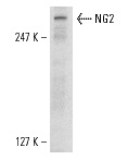
NG2 (F-12) - sc-166179 from Santa Cruz Biotechnology
|
||||||||||
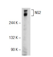
NG2 (G-20) - sc-30923 from Santa Cruz Biotechnology

|
||||||||||
NG2 (L-20) - sc-30921 from Santa Cruz Biotechnology
|
||||||||||
NG2 (H-300) - sc-20162 from Santa Cruz Biotechnology
|
||||||||||
NG2 (G-9) - sc-166251 from Santa Cruz Biotechnology
|
||||||||||
NG2 (7.1) - sc-53508 from Santa Cruz Biotechnology
|
||||||||||
NG2 (LHM 2) - sc-53389 from Santa Cruz Biotechnology
|
||||||||||
NG2 (9.2.27) - sc-80003 from Santa Cruz Biotechnology
|
||||||||||
p-NG2 (Thr 2252) - sc-33038 from Santa Cruz Biotechnology
|
||||||||||
Anti-NG2 antibody - ab83508 from Abcam
|
||||||||||
Anti-NG2 antibody - ab83178 from Abcam
|
||||||||||
Anti-NG2 antibody - ab104535 from Abcam
|
||||||||||
Anti-NG2 antibody - ab129051 from Abcam
|
||||||||||
Anti-NG2 antibody - ab86067 from Abcam

|
||||||||||
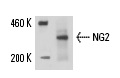
Anti-NG2 antibody [ EPR9195] - ab139406 from Abcam
![Anti-NG2 antibody [ EPR9195] (ab139406) at 1/1000 dilution + A375 cell lysate at 10 µgSecondaryHRP labelled goat anti-rabbit at 1/2000 dilution](http://www.bioprodhub.com/system/product_images/ab_products/2/sub_4/152_NG2-Primary-antibodies-ab139406-1.jpg)
|
||||||||||
Anti-NG2 antibody [LHM 2] - ab20156 from Abcam
|
||||||||||
Anti-CSPG / NG2 Antibody - LS-C79283 from LifeSpan Bioscience
|
||||||||||
Anti-CSPG / NG2 Antibody - LS-C152910 from LifeSpan Bioscience
|
||||||||||
Anti-CSPG4 / NG2 Antibody - LS-C22107 from LifeSpan Bioscience
|
||||||||||
Anti-CSPG4 / NG2 Antibody - LS-C22108 from LifeSpan Bioscience
|
||||||||||
Anti-CSPG4 / NG2 Antibody (clone 9.9.27) - LS-C22105 from LifeSpan Bioscience
|
||||||||||
Anti-CSPG4 / NG2 Antibody - LS-C22112 from LifeSpan Bioscience
|
||||||||||
Anti-CSPG4 / NG2 Antibody - LS-C22106 from LifeSpan Bioscience
|